Download
1 / 31
330 likes | 726 Views
Acute Pancreatitis Arefe Hedayati. Normal Anatomy & Physiology. neutralize chyme digestive enzymes hormones. Exocrine Function. common bile duct. BODY. TAIL. HEAD. pancreatic duct. ampulla. UNCINATE. Digestive Enzymes in the Pancreatic Acinar Cell. PROTEOLYTIC LIPOLYTIC ENZYMES

E N D
Normal Anatomy & Physiology • neutralize chyme • digestive enzymes • hormones
Exocrine Function common bile duct BODY TAIL HEAD pancreatic duct ampulla UNCINATE
Digestive Enzymes in the Pancreatic Acinar Cell • PROTEOLYTIC LIPOLYTIC ENZYMES • ENZYMESLipase • TrypsinogenProphospholipaseA2 • ChymotrypsinogenCarboxylesterase lipase • Proelastase • Procarboxypeptidase A NUCLEASES • Procarboxypeptidase B Deoxyribonuclease (DNAse) • Ribonuclease (RNAse) • AMYOLYTIC ENZYMES • Amylase OTHERS • Procolipase • Trypsin inhibitor
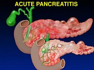
Acute PancreatitisDefinition • Acute inflammatory process involving the pancreas • Usually painful and self-limited • Isolated event or a recurring illness • Pancreatic function and morphology return to normal after (or between) attacks
prematureenzyme activation Acute PancreatitisPathogenesis • acinar cell • injury prematureenzyme activation failed protectivemechanisms
premature enzyme activation autodigestion of pancreatic tissue local vascular insufficiency activation of white blood cells release ofenzymes into the circulation localcomplications distantorgan failure
Acute PancreatitisAssociated Conditions • Pancreas divisum • Hereditary • Hypercalcemia • Viral infections • Mumps • Coxsackievirus • End-stage renal failure • Penetrating peptic ulcer • Cholelithiasis • Ethanol abuse • Idiopathic • Medications • Hyperlipidemia • ERCP • Trauma
Acute PancreatitisCausative Drugs • AIDS therapy:didanosine, pentamidine • Anti-inflammatory:sulindac, salicylates • Antimicrobials: metronidazole, sulfonamides, tetracycline, nitrofurantoin • Diuretics: furosemide, thiazides • IBD: sulfasalazine, mesalamine • Immunosuppressives: azathioprine, 6-mercaptopurine • Neuropsychiatric:valproic acid • Other: calcium, estrogen, tamoxifen, ACE-I
Acute PancreatitisPathogenesis SEVERITY Mild Severe • STAGE 1: Pancreatic Injury • Edema • Inflammation • STAGE 2: Local Effects • Retroperitoneal edema • Ileus • STAGE 3: Systemic Complications • Hypotension/shock • Metabolic disturbances • Sepsis/organ failure
Acute PancreatitisClinical Presentation • Abdominal pain • Epigastric • Radiates to the back • Worse in supine position • Nausea and vomiting • Fever
Acute PancreatitisDifferential Diagnosis • Choledocholithiasis • Perforated ulcer • Mesenteric ischemia • Intestinal obstruction • Ectopic pregnancy
Acute PancreatitisDiagnosis • Symptoms • Abdominal pain • Laboratory • Elevated amylase or lipase • > 3x upper limits of normal • Radiology • Abnormal sonogram or CT
Acute PancreatitisClinical Manifestations PANCREATIC PERIPANCREATIC SYSTEMIC Mild: edema, inflammation, fat necrosis Severe:phlegmon, necrosis, hemorrhage, infection, abscess, fluid collections Retroperitoneum, perirenal spaces, mesocolon, omentum, and mediastinum Adjacent viscera: ileus, obstruction, perforation Cardiovascular: hypotension Pulmonary: pleural effusions, ARDS Renal: acute tubular necrosis Hematologic: disseminated intravascular coag. Metabolic:hypocalcemia, hyperglycemia
Acute PancreatitisDiagnosis • EtOH: history • Gallstones: abnormal LFTs & sonographic evidence of cholelithiasis • Hyperlipidemia: lipemic serum, Tri>1,000 • Hypercalcemia: elevated Ca • Trauma: history • Medications: history, temporal association
Severity Scoring Systems • Ranson and Glasgow Criteria (1974) • based on clinical & laboratory parameters • scored in first 24-48 hours of admission • poor positive predictors (better negative predictors) • APACHE Scoring System • can yield a score in first 24 hours • APACHE II suffers from poor positive predictive value • APACHE III is better at mortality prediction at > 24 hours • Computed Tomography Severity Index • much better diagnostic and predictive tool • optimally useful at 48-96 hours after symptom onset
Ranson Criteria AT ADMISSION • Age > 55 years • WBC > 16,000 • Glucose > 200 • LDH > 350 IU/L • AST > 250 IU/L WITHIN 48 HOURS • HCT drop > 10 • BUN > 5 • Arterial PO2 < 60 mm Hg • Base deficit > 4 mEq/L • Serum Ca < 8 • Fluid sequestration > 6L
RansonCriteria AT ADMISSION • Age > 70 years • WBC > 18,000 • Glucose > 220 • LDH > 400 IU/L • AST > 250 IU/L WITHIN 48 HOURS • HCT drop > 10 • BUN > 2 • Base deficit > 5 mEq/L • Serum Ca < 8 • Fluid sequestration > 4L
Severe Acute Pancreatitis • Scoring systems • 3Ranson criteria • 8 APACHE II points • 5 CT points • Organ failure • shock (SBP < 90 mmHg) • pulmonary edema / ARDS (PaO2 < 60 mmHg) • renal failure (Cr > 2.0 mg/dl) • Local complications • fluid collections pseudocysts • necrosis (mortality 15% if sterile, 30-35% if infected) • abscess
Treatment of Mild Pancreatitis • Pancreatic rest • Supportive care • fluid resuscitation – watch BP and urine output • pain control • NG tubes and H2 blockers or PPIs are usually not helpful • Refeeding(usually 3 to 7 days) • bowel sounds present • patient is hungry • nearly pain-free (off IV narcotics) • amylase & lipase not very useful here
Treatment of Severe Pancreatitis • Pancreatic rest & supportive care • fluid resuscitation* – may require 5-10 liters/day • careful pulmonary & renal monitoring – ICU • maintain hematocrit of 26-30% • pain control – PCA pump • correct electrolyte derangements (K+, Ca++, Mg++) • Rule-out necrosis • contrasted CT scan at 48-72 hours • prophylactic antibiotics if present • surgical debridement if infected • Nutritional support • may be NPO for weeks • TPN vs. enteral support (TEN)
1-clinical manifestation 2-lypase/amylase 3-sonography Severity 1-manifestation 2-ranson or APACHE score 3-CT MILD SEVER SUPPORTIVE CARE ICU
ICU Antibiotic Supportive care FNA remission SEPSIS/INFLAMATION debridmane
Conclusions • Acute pancreatitis is a self-limited disease in which most cases are mild. • Gallstones and alcohol are the leading causes of acute pancreatitis. • In mild pancreatitis, nutritional support is usually not required • In severe pancreatitis, nutritional support will likely be required with the enteral route preferred over TPN because of both safety and cost.