Download
1 / 10
0 likes | 13 Views
Dr. Shaji is renowned for his expertise in CBCT scanning, providing patients with cutting-edge diagnostic imaging services.

E N D
The Role of CBCT in Detecting and Diagnosing Oral Pathologies
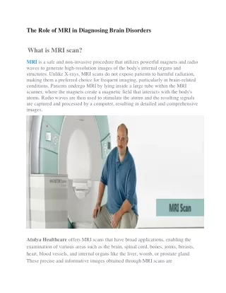
Introduction to CBCT Cone Beam Computed Tomography (CBCT) is a cutting-edge imaging technology widely used in dentistry and oral health care. Unlike traditional 2D X-rays, CBCT creates highly detailed 3D images by emitting a cone-shaped X-ray beam that rotates around the patient. The data captured from multiple angles is processed into high-resolution, three-dimensional images, offering a more comprehensive view of the teeth, bones, soft tissues, and surrounding structures. The main advantage of CBCT lies in its ability to provide detailed, clear, and accurate information, which is crucial for diagnosing a variety of oral and maxillofacial conditions. It allows for better visualization of structures such as the teeth, jawbone, sinuses, nerves, and surrounding tissues, leading to more precise treatment planning. CBCT is particularly valuable in areas like dental implant planning, orthodontics, pathology detection, and the evaluation of complex dental conditions such as cysts, tumors, or bone fractures. CBCT imaging has revolutionized the field of dentistry by enhancing diagnostic accuracy, improving surgical outcomes, and minimizing the need for invasive procedures. With its ability to capture 3D data, CBCT has become an indispensable tool for dental professionals, helping them provide more personalized and effective patient care.
Oral Pathologies Overview Oral pathologies refer to diseases or conditions that affect the tissues of the mouth, jaw, and surrounding structures. Identifying these conditions early is critical to providing effective treatment and improving patient outcomes. Here’s an overview of common oral pathologies:CBCT can detect caries in hard-to-see areas, such as between teeth or under restorations, with greater accuracy than 2D X-rays. 1. Dental Caries (Tooth Decay) • Description: • Dental caries is the destruction of tooth structure due to the activity of bacteria that feed on sugars and produce acids. It often leads to cavities or holes in the teeth. • Signs & Symptoms: Tooth sensitivity, visible holes or pits, pain when chewing or drinking hot/cold beverages. • Role of CBCT: CBCT can detect caries in hard-to-see areas, such as between teeth or under restorations, with greater accuracy than 2D X-rays. 2. Periodontal Disease (Gum Disease) • Description: • Periodontal disease involves inflammation and infection of the gums, ligaments, and bones supporting the teeth. It can progress from gingivitis (early-stage) to periodontitis (severe stage), leading to tooth loss if untreated. • Signs & Symptoms: Swollen, bleeding gums, bad breath, receding gums, and loose teeth. • Role of CBCT: CBCT allows for 3D visualization of bone loss around the teeth, providing precise data for treatment planning and surgical interventions.
3. Oral Cysts • Description: • Cysts are fluid-filled sacs that can develop in the oral cavity or jaw. Common types include dentigerous cysts, periapical cysts, and radicular cysts. • Signs & Symptoms: Swelling or lump in the mouth, jaw pain, or discomfort. • Role of CBCT: CBCT helps in identifying the size, location, and relationship of cysts to surrounding structures, aiding in treatment and surgical planning. 4. Oral Tumors and Neoplasms • Description: • Oral tumors may be benign (non-cancerous) or malignant (cancerous). Examples include squamous cell carcinoma, salivary gland tumors, and fibromas. • Signs & Symptoms: Lumps or masses in the mouth, persistent sores that don’t heal, unexplained pain, or difficulty swallowing. • Role of CBCT: CBCT helps detect tumors early, providing detailed imaging that supports the diagnosis and guides the surgical removal of abnormal growths. 5. Jaw Fractures and Trauma • Description: • Jaw fractures result from trauma or accidents, such as falls or car accidents, and can affect the lower jaw (mandible) or upper jaw (maxilla). • Signs & Symptoms: Pain, swelling, difficulty moving the jaw, or a misaligned bite. • Role of CBCT: CBCT provides a precise 3D view of fractures and bone displacement, allowing for accurate diagnosis and planning of treatments like splints or surgery.
What is CBCT? Cone Beam Computed Tomography (CBCT) is a specialized type of X-ray imaging technique used primarily in dentistry and other medical fields to create detailed, three-dimensional images of the teeth, jaw, and surrounding structures. Unlike traditional 2D X-rays, which produce flat images, CBCT produces high-resolution 3D images that allow for a more comprehensive view of the oral and maxillofacial region. How Does CBCT Work? • Cone-Shaped X-Ray Beam: CBCT uses a cone-shaped X-ray beam that rotates around the patient, capturing multiple images from different angles. • Data Reconstruction: The collected data is then processed by sophisticated software to create a detailed 3D image of the area being scanned, such as the teeth, bones, soft tissues, and other structures. • High-Resolution Images: CBCT offers superior image quality compared to traditional 2D X-rays, providing clear and precise views of the hard and soft tissues in the area of interest.
Key Features of CBCT: 3D Imaging • Description: CBCT produces detailed three-dimensional images of the teeth, jaw, and surrounding structures, unlike traditional 2D X-rays. It offers a comprehensive view of the entire area being scanned, allowing for better evaluation of complex anatomy. • Benefit: Helps detect conditions that might be missed in 2D images, like hidden cavities, fractures, or lesions. High-Resolution Images • Description: CBCT provides high-resolution, detailed images, allowing dental professionals to see minute details such as small fractures, cysts, or tumors within the bone and soft tissues. • Benefit: Enables accurate diagnosis and precise treatment planning, ensuring better outcomes for dental procedures like implants or extractions.
Lower Radiation Exposure • Description: Compared to conventional CT scans, CBCT uses a significantly lower radiation dose while still providing high-quality images. • Benefit: Reduces the risks associated with radiation exposure, making it safer for patients, especially for those requiring multiple scans. Faster Imaging Process • Description: CBCT scans are relatively quick to perform, typically taking only a few minutes to complete. • Benefit: Minimizes patient discomfort and allows for efficient diagnosis and treatment planning. Non-invasive and Painless • Description: The CBCT scan is a non-invasive procedure, requiring no surgical incisions or injections. The patient simply sits in a chair while the scanner captures the necessary images. • Benefit: Ensures a comfortable experience for patients, making it ideal for a wide range of age groups, including children. Ability to Visualize Hard and Soft Tissues • Description: CBCT is capable of capturing detailed images of both hard tissues (such as teeth and bone) and soft tissues (such as nerves, blood vessels, and mucous membranes). • Benefit: Provides a comprehensive view of the oral and maxillofacial region, aiding in accurate diagnosis and treatment of both dental and non-dental issues.
Identifying Cysts and Tumors with CBCT Cone Beam Computed Tomography (CBCT) is a powerful diagnostic tool for identifying cysts, tumors, and other abnormal growths in the oral and maxillofacial regions. The high-resolution 3D images provided by CBCT allow dental professionals to precisely locate, measure, and assess the size, shape, and relationship of these lesions to surrounding structures. Here’s how CBCT aids in the identification of cysts and tumors: 1. High-Resolution Imaging for Accurate Detection • Detailed 3D Views: CBCT scans create high-quality 3D images that provide detailed views of the bone, teeth, and soft tissues. This level of detail helps identify even small cysts or tumors that might not be visible on traditional 2D X-rays. • Benefit: Accurate detection of cysts and tumors at an early stage, leading to quicker diagnosis and treatment. 2. Clear Visualization of Lesions • Cyst Identification: CBCT can clearly distinguish cysts, which are fluid-filled sacs, from other types of lesions, providing valuable information about their location, size, and impact on adjacent teeth and bone. • Tumor Detection: CBCT is also effective in detecting both benign and malignant tumors. It provides essential information on the borders of the lesion, the extent of bone involvement, and the involvement of nearby vital structures (e.g., nerves, blood vessels).
Accurate Measurement and Planning for Treatment • Precise Sizing: CBCT offers precise measurements of cysts and tumors, including their size, shape, and orientation. This information is critical when planning surgical procedures, such as tumor resection or cyst removal. • Surgical Planning: CBCT helps plan the surgical approach by showing the relationship of the lesion to critical anatomical structures such as nerves, blood vessels, and the maxillary sinus. It assists in planning the safest, most effective surgical route. • Benefit: Reduces surgical risks and ensures more accurate excision of the lesion. 4. Differentiation of Lesion Types • Benign vs. Malignant Lesions: CBCT can help distinguish between benign cysts (such as dentigerous cysts) and more aggressive tumors (like ameloblastomas or squamous cell carcinoma). This differentiation is crucial for deciding on the treatment approach. • Benefit: Helps avoid misdiagnosis and ensures appropriate treatment for the specific type of lesion.
Conclusion: In conclusion, Dr. Shaji's expertise in performing CBCT scans makes him one of the best in the field. With a deep understanding of the technology and its applications, Dr. Shaji ensures that every patient receives the highest quality diagnostic imaging, enabling accurate detection, effective treatment planning, and optimal outcomes. Whether you're seeking to identify oral pathologies, assess bone structure, or plan for dental implants, Dr. Shaji's advanced CBCT imaging services provide the clarity and precision needed for exceptional care. Trusting Dr. Shaji with your CBCT scan means choosing a reliable, skilled professional who prioritizes your health and well-being at every step. Dr. Shaji is renowned for his expertise in CBCT scanning, providing patients with cutting-edge diagnostic imaging services. With a commitment to precision and accuracy, Dr. Shaji leverages the latest CBCT technology to deliver clear, detailed 3D images that are crucial for identifying oral pathologies, planning treatments, and ensuring optimal outcomes. His deep understanding of both the technology and its clinical applications ensures that every scan is conducted with the highest level of care and expertise. For those seeking accurate diagnostics and personalized treatment planning, Dr. Shaji’s CBCT services stand out as a top choice for exceptional dental care.