Download
1 / 5
70 likes | 195 Views
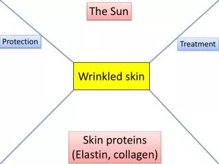
Discover the unique functions and structures of fibrous proteins like collagen, elastin, and keratin. Learn about their roles in the body and how they contribute to skin, connective tissues, and more. Dive into the mechanical properties and amino acid compositions that make these proteins essential for our bodies.

E N D
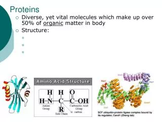
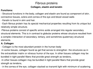
Fibrous proteins • Collagen, elastin and keratin • Functions: - Structural functions in the body, collagen and elastin are found as component of skin, connective tissues, sclera and cornea of the eye and blood vessel walls - Keratin is found in skin and hair. - Each fibrous protein has its special mechanical properties resulting from its unique but relatively simple structure. • Fibrous proteins consist of specific amino acids arranged into regular secondary structural elements. This is in contrast to globular proteins whose structure resulted from a complex interaction of secondary, tertiary, and sometimes quaternary structural elements. • A. Collagen • - Collagen is the most abundant protein in the human body • In some tissues, collagen found as gel that serves to strengthen the structures as in the extracellular matrix or vitreous humor of the eye. In other tissues collagen may be bundled in tight parallel fibers that provide great strength as tendons, • In other tissues collagen may be bundled in tight parallel fibers that provide great strength as tendons, • in the cornea of the eye, collagen stacked so transmit light with minimum of scattering.
- Collagen in the bone occurs as fibers arranged at an angle to each other so as to resist mechanical shear from any direction Structure of collagen Types of collagen Collagen is formed from three polypeptides called α-chains which wrap around each other in a triple helix forming a rope-like structure. The three polypeptide chains are held together by H-bonds Variations in amino acids sequence of the α-chains result in structural components that are in the same size (around 1000 amino acids) but slightly with different properties. The α-chains are combined to form the various types of collagen found d in tissue. Figure 3.23
Amino acid sequence: • the primary structure of collagen is unusual in that glycine is found in every third position of the polypeptide chain, the glycine residue is a part of a repeating sequence –Gly-X-Y where X is frequently is proline and Y often hydroxyproline or hydroxylysine • Triple-helical structure • Collagen doesn't fold into a compact structure • - It has an elongated triple-helical structure , the amino acid side chains are placed outside of the molecule. • This allow the interaction between triple-helical molecules that lead to aggregation of collagen monomers into long fiber • Hydroxyproline and hydroxylysine: • These amino acids are rarely found in other proteins and found extensively in collagen • They resulted from hydroxylation of Proline and Lysine after their incorporation into polypeptide chains. (Posttranslation modification) • Hydroxproline is important in stabilizing the triple-helical structure • Glycosylation • The hydroxyl group of the hydroxylysine residues of collagen maybe glycosylated. Most commonly glucose and galactose.
Elastin • Elastin is a connective tissue protein with a rubber like properties. Elastin fibers like are found at lungs, wall of blood vessels and elastic ligaments. Elastin can be stretched to several times their normal length but recoil to their original shape when the stretching force is relaxed • Structure of elastin • Amino acid composition • Elastin is composed primarily of small, non-polar amino acid residues as glycine, alanine and valine. • Elastin also rich in lysine and proline but little hydroxyproline and no hydroxylysine. • Interchain cross-link • Elastin fiber are formed as three dimensional network of cross-linked polypeptide that have an irregular conformation. • The cross-link involve lysine. 4 lysine residue from 4 separate chains can be covalently joined to produce a desmosine cross link results in inter-connected, rubbery network that can stretch and bend in any direction when stressed giving connective tissue its elasticity.
α-Keratins • The α-Keratins are proteins that form tough fibers. They are found in hair, nails and outer epidermal layer of mammals. • α-Keratins are also constituents of intermediate filaments of eth cytoskeleton in certain cells. • α-Keratins are rich in cysteine covalent disulfide cross-links between adjacent polypeptide chains thus producing fibers that insoluble and resistant to stretching. • The α-Keratins of hair is an example of a protein constructed almost of α-helices. • Hair is composed of dead cells. Each cell is packed with keratin macrofibrils • Macrofibriles are formed of microfibril embedded into a protein matrix. Each microfibril is formed from protofibrils. • Protofibrils are formed from α-helix protein