Esophageal Motility Disorders
450 likes | 1.45k Views
Esophageal Motility Disorders. Iskander Al- Githmi , MD, FRCSC, FRCSC (Ts & CDS), FACS, FCCP Consultant & Asst. Professor of Cardiothoracic Surgery King Abdulaziz University College of Medicine. Anatomy of The Esophagus.

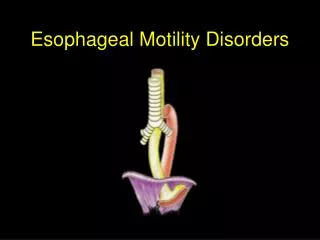
Esophageal Motility Disorders
E N D
Presentation Transcript
Esophageal Motility Disorders Iskander Al-Githmi, MD, FRCSC, FRCSC (Ts & CDS), FACS, FCCP Consultant & Asst. Professor of Cardiothoracic Surgery King Abdulaziz University College of Medicine
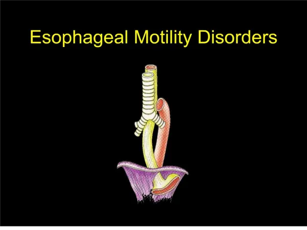
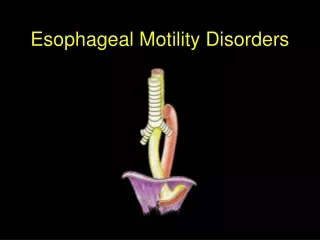
Anatomy of The Esophagus • The esophagus is a hollow muscular organ, approximately 25cm in length that extend from the pharynx to the stomach • The pharynx is a muscular tube, approximately 12cm in length that serve as entry to the esophagus and respiratory tract.
Anatomy of The Esophagus • Cervical Esophagus: Just lies to the left of midline behind the larynx and the trachea. The entry to esophagus called upper esophageal sphincter (UES). • Thoracic Esophagus: The upper part passes behind the carina & Lt. main stem bronchus. The lower part passes behind the left atrium. • Abdominal Esophagus: Is the smallest portion of the esophagus (2-4cm length). It has lower esophageal sphincter (LES)- non anatomical with normal resting pressure 10-20mmHg.
Anatomy of The Esophagus • Normal esophageal narrowing: • UES at the level of cricoid cartilage 14mm in diameter. • Broncho-aortic constriction 17mm in diameter. • LES (19mm) as it travels the diaphragm & located 3-5cm at distal part of the esophagus. • Clinical Importance of normal esoph. narrowing: • Potential for development of diverticulum's (Zenker) in the neck. • Potential for perforation during esophagoscopy • Pills-induced stricture.
Anatomy of The Esophagus • The esophageal wall: • The proximal esophagus is predominantly striated muscle. • The distal esophagus is predominantly smooth muscle. • The mid esophagus contained a graded transition of striated and smooth muscle.
Anatomy of The Esophagus The esophageal wall: • The muscle oriented in two perpendicular opposing layers an inner circular layer and outer longitudinal layers both called muscularis propria. • The outermost layer of the esophagus called adventitia (fibro-areolar layer), but no serosa. This may contribute for cancer spread. • Underneath the adventitia there is a longitudinal muscle layer and beneath there is circular layer. • Between the two muscle layers there are network of sympathetic and parasympathetic fibers (myentric plexus)
Anatomy of The Esophagus • The esophageal wall: • Beneath the muscle layers lies the submucosa which contain mucus gland, blood and lymphatic vessels and network works of nerve fibers (meissners). • Beneath the submucosa is the mucosa which consist of squamous epithelium except the distal 2cm at G-E junction (Z-line) or transition to columnar epithelium.
Anatomy of The Esophagus • Blood supply & venous drainage • Cervical esophagus received its arterial blood from inferior thyroid artery. • Thoracic esophagus received its arterial blood from bronchial, aorta, left gastric artery and from inferior phrenic artery. • The esophageal veins drain to periesophageal venous network & to inferior thyroid vein in the neck and to azygos and hemiazygos veins in the thorax.
Anatomy of The Esophagus • Lymphatic drainage: • The lymphatic plexus are located in the mucosa and the muscular layers drained to mediastinal lymph nodes. Clinical facts about the esophagus • Cervical esophagus is 5 cm in length and 15cm distance from upper incisors • Thoracic esophagus is 12cm in length and 25cm distance from upper incisors • Lower esophagus is 2cm in length & 38cm from upper incisors
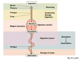
Physiology of The Esophagus • The function of the esophagus is to transport the ingested material from the pharynx to the stomach by peristaltic waves. • Primary peristalsis: Triggered by the swallowing center in the brain stem and the contraction wave travel at speed 2cm/s. • Secondary peristalsis: Induced by esophageal distension from retained bolus, refluxed material. Its role is to clear the esophagus form retained bolus.
Physiology of The Esophagus • Tertiary peristalsis: Are non peristaltic contraction and play no known physiological role. Frequently observed in elderly people called (presbyesophagus), also seen in motility disorders.
Physiology of The Esophagus • Mechanism of swallowing • During the pharyngeal phase of swallowing, a primary peristalsis is created, that relax the UES and forces the food bolus through it. The UES remain constricted and has resting pressure of 20-60 mmHg. The peristaltic waves travel at the speed 2cm/s and reach the stomach in 5-10 second
Physiology of The Esophagus • Secondary peristalsis get initiated if the primary peristalsis failed to get food to the stomach and the esophagus became distended.
Esophageal Motility Disorders • Achalasia • Spastic esophageal motility disorders such as diffuse esophageal spasm, nutcracker esophagus and hypertensive LES • Secondary esophageal motility disorders related to scleroderma, diabetes, alcohol consumption ..
Esophageal Motility Disorders • Achalasia (failure to relax) • Is the only esophageal motility disorder with an established pathology. • The predominant pathophysiology of achalasia is the loss of Auerbach ganglion cells from the wall of the esophagus ,starting at LES and progress proximally. • Incidence is 1-3 / 100,000 population / year.
Esophageal Motility Disorders • Achalasia (failure to relax) • Characterized by failure of LES to relax completely during swallowing • The loss of nerve ganglion along the esophageal wall cause a peristalsis leading to stasis of food and subsequent dilatation. • Manometry may reveal elevated LES pressure > 40 mmHg in 60% of patients.
Esophageal Motility Disorders • Spastic esophageal motility disorders • Diffuse esoph.spasm (DES): This is probably related to fragmental degeneration of vagal nerve fibers. • Characterized by simultaneous, repetitive high pressure muscular contraction within the esophagus. • The muscular wall is thickened, hypertrophied and is hypersensitive to stretching.
Esophageal Motility Disorders • Scleroderma esophagus • Collagen vascular disease. • Characterized by smooth muscle hypertrophy and mainly involve the distal 2/3 of esophagus gradually lead to loss of peristalsis and weakening of LES causing GERD. • Involve the esophagus in 80% of patient with scleroderma.
Esophageal Motility Disorders • Clinical History • Achalasia: • The hall mark is dysphagia to both solid and liquid. • Regurgitation commonly occur at night • Retrosternal chest pain. • Heartburn occur in 30% of patients which may be related to food fermentation and lactic acid.
Esophageal Motility Disorders • Clinical History • Spastic motility disorders • Chest pain is the hall mark which may mimic angina due to esophageal distension. • Dysphagia to both solid and liquid. • Scleroderma • Involve the esophagus in 80% of patients. • Symptoms are related to GERD [dysphagia, heartburn and regurgitation].
Esophageal Motility Disorders • Problems to be considered • Coronary Artery Disease (CAD). • Mechanical obstruction (tumor). • Achalaisa and scleroderma increase risk of esophageal cancer.
Esophageal Motility Disorders • Diagnosis • History • Physical examination-unremarkable • Barium Swallow Bird peak appearance- classic for achalasia Rosary beads or corkscrew- classic for DES
Esophageal Motility Disorders • Diagnosis • Esophagoscopy to rule out tumor or inflammatory lesion but not to diagnose esophageal dysmotility. • Manometry study is to evaluate the esophageal motor pattern, contraction amplitude and LES pressure.
Esophageal Motility Disorders • Treatment • The primary goal is symptomatic relief directed at relieving the physiologic obstruction at the level of LES by surgical or balloon dilatation. • Nitrate and Ca channel & B blockers are currently used in all patients with esophageal motility disorders. • Antireflux therapy e.g proton pump inhibitors (esomeprazol) + prokinetic such as motilium or erythromycin.
Esophageal Motility Disorders • Treatment • Botulinum toxin injection (Botox): Injected edoscopically in 4 quadrants into LES in treating patient with achalasia. Botox is a potential inhibitor of acetylcholine release from nerve terminals. It is indicated in those pt. not candidate for surgery or refuse surgery. • Endoscopic balloon dilatation: This is the standard therapy for patients with achalasia. • The mechanism based on disruption of circular muscle. • Balloon dilatation response rate is 70%
Esophageal Motility Disorders • Treatment • Surgery (Heller Myotomy): surgical treatment targets to disrupt the LES. • This can be performed thoracoscopic or laparascopic. • Outcome is excellent 80-100% response rate.
Barrett Esophagus Definition: Intestinal metaplasia Risk factors Age Male GERD Smoking Treatment: Antireflux therapy Medical: Pump inhibitors (esomeprazole) Prokinetic meds (Motilium) Annual Surveillance (esophagoscopy) Surgical: Fundoplication + Annual Surveillance Complications: Dysplasia Adenocarcinoma <1%/Yr