Download

1 / 19
200 likes | 337 Views
Explore the intricacies of embryology and congenital anomalies of the female genital system, covering key components such as gonads, duct systems, and external genitalia. Learn about the differentiation of sexes, formation of gonads, and abnormalities that can occur in the ovaries. Discover the fascinating process of external genitalia development and the various anomalies that may arise. Get insights into the complexities of female genital duct formation and the interplay with gonadal development. Don't miss this informative presentation by Dr. Manal Beheiry from Zagazig University, Egypt.

E N D
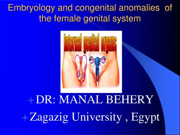
Embryology and congenital anomalies of the female genital system • DR • DR: MANAL BEHERY • Zagazig University , Egypt
C Components which form female and male reproductive systems are: 1. Gonads Ovaries or testes 2. Genital Duct Systems Mesonephric and Paramesonephric ducts 3. External Genitalia
Genotype of embryo 46XX or 46XY is established at fertilization • SRY (sex-determining region on Y) that encodes a protein called testis-determining factor (TDF) are responsible for male differentiation
Indifferent Embryo • Weeks 1-6 sexually indifferent or undifferentiated stage • Week 7 begins phenotypic sexual differentiation • Week 12 female or male characteristics of external genitalia can be recognized and completed at 20 weeks.
At 4TH Week of gestation • Mesonephric Duct • extending from the • mesonephros • (Wolff’s body) • to the cloaca • (urogenital sinus)
A Swelling on either side of dorsal mesentry on medial side of mesonephric duct forms the urogenital ridge At 5TH Week of gestation
Primitive germ cells migrate from yolk sac through dorsal mesentry to reach genital ridge These germ cells stimulate coelomic epithelium and underlying mesoderm to proliferate and form primitive sex cords Formation and Differentiation of Gonads
At 6TH week gestation Paramesonephric or Mullerian Duct develops lateral to the Mesonephric ”wolffian “ Duct
The middle and caudal parts of the Mullerian ducts undergoes medial migration and fusion. • The cranial 1/3 → tubes. • The middle 1/3 → uterus and cervix. • Caudal 1/3 → upper 3/4 of vagina.
2 main Principle • Internal genital organs develop in close association with urinary tract So gross malformation of uterus and tube are commenaly associated with anomalies of kidney and ureter. • Development of gonads is separt from that of the ducts So functional ovary are usually present when uterus, vagina are absent
In ovary the absence of testosterone inhibits the development of the mesonephric ducts. The atretic remains form the epoophoron, paraoophoron and Gartner’s ducts. In absence of AMH, paramesonephric ducts form the female internal genital tract. Female Genital Duct Formation
Abnormalities of the ovaries: • 1) agenesis or complete absence. • 2) Gonadal dysgenesis "streak gonads" as in Turner syndrome. • 3) Failure of descent into the pelvis. • 4) Ovotestis “true hermaphrodite” In which combined ovarian and testicular tissues seen.
Development external genitalia • Early, similar in both sexes • 6th wk, three external protuberance surround cloacal membrane, the left and right genital swellings meet anteriorly to form the genital tubercle. • 12th wk identify difference. • Genital swelling labioscrotal folds scrotum or labia major • Genital tubercle phallus penis or clitoris
Interesting, right? This is just a sneak preview of the full presentation. We hope you like it! To see the rest of it, just click here to view it in full on PowerShow.com. Then, if you’d like, you can also log in to PowerShow.com to download the entire presentation for free.