Download

1 / 33
380 likes | 835 Views
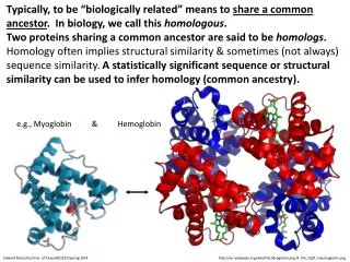
Structures of Myoglobin and Hemoglobin. Myoglobin ( Mb ) - monomeric protein that facilitates the diffusion of oxygen in vertebrates Hemoglobin ( Hb ) - tetrameric protein that carries oxygen in the blood

E N D
Structures of Myoglobin and Hemoglobin • Myoglobin (Mb) - monomericprotein that facilitates the diffusion of oxygen in vertebrates • Hemoglobin (Hb) - tetramericprotein that carries oxygen in the blood • Heme consists of a tetrapyrrole ring system called protoporphyrin IX complexed with iron • Heme of Mb and Hb binds oxygen for transport
Protein component of Mb and Hb is globin • Myoglobin is composed of 8 a helices • Heme prosthetic group binds oxygen • His-93 is complexed to the iron atom, and His-64 forms a hydrogen bond with oxygen • Interior of Mb almost all hydrophobic amino acids • Heme occupies a hydrophobic cleft formed by threea helices and two loops
Hemoglobin (Hb) • Hb is ana2b2tetramer (2aglobin subunits, 2bglobin subunits) • Each globin subunit is similar in structure to myoglobin • Each subunit has a heme group • Theachain has7ahelices,bchain has 8ahelices
Hemoglobin tetramer (a) Human oxyhemoglobin (b) Tetramer schematic
Oxygen Binding to Mb and Hb • Oxymyoglobin - oxygen bearing myoglobin • Deoxymyoglobin - oxygen-free myoglobin • In oxymyoglobin, six ligands are coordinated to the ferrous ion in octahedral symmetry • Oxygen is coordinated between the iron and the imidazole sidechain of His-64
Oxygen-binding site of whale oxymyoglobin • Octahedral geometry of coordination complex (six ligands around iron) • His-93 (proximal histidine) liganded to Fe • His-64 (distal histidine)
Oxygen-binding curves (a) Comparison of O2-binding to Mb and Hb
Oxygen-Binding Curves of Myoglobin and Hemoglobin • Curves show reversiblebinding of O2 to Mb and Hb • Fractionalsaturation (Y) is plotted versus the partial pressure of oxygen, pO2 (oxygen concentration) • The shape of the Hb curve shows a positivecooperativity in the binding of 4 O2 molecules (i.e. the O2 affinity of Hb increases as each O2 molecule is bound)
Oxygen-binding curves (a) Comparison of O2-binding to Mb and Hb
O2 binding curves (continued) Mb-O2 binding curve is hyperbolic, indicating a single equilibrium constant for binding O2 Hb-O2 binding curve is sigmoidal, and reflects the binding of 4 molecules of O2, one per each heme group
Oxygen-binding curves (a) Comparison of O2-binding to Mb and Hb
Oxygen-binding curves Binding of the R (high-affinity) and T (low affinity) forms of Hb
Conformational changes in a hemoglobin chain induced by oxygenation • Oxygen binding to Fe pulls the His toward ring plane • Helix with His shifts position, disrupting some ion pairs between subunits (blue to red position)
Oxygen-binding site of whale oxymyoglobin • Octahedral geometry of coordination complex (six ligands around iron) • His-93 (proximal histidine) liganded to Fe • His-64 (distal histidine)
Hemoglobin is an Allosteric Protein • Oxygen binding and release from Hb are regulated by allosteric interactions • Allosteric effectors (modulators) bind to a protein at a site separate from the functional binding site (may be activators or inhibitors) • The activity of an allosteric protein is regulated by allosteric effectors
Two conformations of hemoglobin: T and R • Active (R state) and inactive (T state) forms are in rapidequilibrium in allosteric proteins • Binding of substrates and allosteric activators stabilize the R state and shift the equilibrium in the Rdirection • Allosteric inhibitors stabilize the T state and shift the equilibrium in the Tdirection
Bisphospho-D-glycerate (2,3BPG) • 2,3BPG is an allosteric effector of Hb • 2,3BPG lowers the affinity of deoxyHb for oxygen (raises the P50 of Hb from ~12 to ~26 torr) • Negatively charged 2,3BPG is bound to six (+) charged groups of deoxyhemoglobin
Binding of 2,3BPG to deoxyhemoglobin • (-) Charges on 2,3BPG pair with (+) charges lining the central cavity, stabilizing the DeoxyHb form • a-Subunits pink,b-subunits blue, heme groups red
Oxygen-binding curves (a) Comparison of O2-binding to Mb and Hb
Bohr effect • Lowering the pH decreases the affinity of Hb for oxygen
Carbamate adduct • Carbon dioxide is transported from the tissues to the lungs in two ways:(1) Dissolved bicarbonate ions(2) Carbamate adducts of hemoglobin (N-terminal globin residues react with CO2 to form carbamates)
Review of Relevant Parameters • Low P50 indicates high O2 affinity • (2) Low pH (through CO2 intake) stabilizes 2,3-BPG and lowers O2 affinity • (3) Raising P50 causes unloading of O2
Case Studies Shock victims are given intravenous HCO3- Why? HCO3- generatesCO2 to the tissues and lowers the O2 affinity of Hb, thus releases O2 from HbO2 to the tissues
Case Studies Fetal Hb (HB-F) contains ser in place of the cationic his at position 143 of the b chains of adult Hb (HB-A). Residue 143 faces the central cavity between the b chains Outcomes: his in Hb-A is protonated and thus binds more tightly to negatively charged 2,3-BPG; ser in Hb-F is not protonated and does not bind to 2,3-BPG as strongly; thus Hb-F has a greater fraction of HbO2
Case Studies Fetal Hb (HB-F) contains ser in place of the cationic his at position 143 of the b chains of adult Hb (HB-A). Residue 143 faces the central cavity between the b chains Outcomes: Hb-F has a greater fraction of HbO2 which means greater O2 affinity and lower P50 (18 torr) Since average P50 for Hb-A is 26 torr, oxygen can efficiently be transferred from maternal blood to fetus
Antibodies Bind Specific Antigens • Vertebrate immune systems synthesize protein antibodies(immunoglobulins) to eliminate bacteria, viruses, other foreign substances • Antibodies specifically recognize and bind antigens • Antibodies are synthesized by lymphocytes (white blood cells)
Heavy chains (blue) and light chains (red) • Disulfide bonds (yellow) • Variable domains colored darker
Stereo view of the immunoglobin fold • Two antiparallelbsheets linked by nonrepetitive segments