Download

1 / 10
100 likes | 189 Views
Explore the use of PtRu5C(CO)16 as a precursor for carbon-supported [PtRu5] nanoparticles. Characterize resulting alloy phases through X-ray spectroscopy and electron microscopy. Details include in situ EXAFS data collection at 673K.

E N D
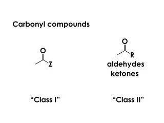
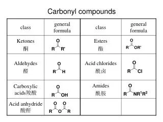
? CO + CH4 H2 [PtRu5] Carbon Black Carbon Black 673K, 1h ca. 200 m2/g Heterometallic Carbonyl Cluster Precursors • Heterometallic molecular cluster precursor • - mediate transport and growth of nanoscale bimetallic particles • Use PtRu5C(CO)16 as a precursor for carbon-supported [PtRu5] nanoparticles • Characterize: Microstructure of the resulting nanometer sized alloy phases • - X-ray spectroscopy • - electron microscopy
Figure 2. Catalyst cell for in-situ EXAFS data collection Figure 1. Si(111) cyrstal bender (D. Adler) Figure 3. Experimental set-up in the X16C “hutch” for in situ X-ray absorption spectroscopy. Includes in situ catalyst cell, gas supply manifold, x-ray detectors, andx-y-z translator.
Experimental Details After depositing and activating the cluster precursor (1h under H2), in situ extended X-ray absorption fine structure (EXAFS) data was collected on the X16C beamline (Scheme 2) at the National Synchrotron Light Source at Brookhaven National Laboratory, Upton, NY (Fig. 1). The beamline utilized a state-of-the-art focusing crystal and catalyst cell (Fig. 2-3). Scanning transmission electron microscopy (STEM) experiments were carried out on a Vacuum Generators HB501 located at the Center for Microanalysis of Materials at the Materials Research Laboratory, Urbana, IL.
20 nm Electron Microscopy On the right is a sample dark field micrograph of [PtRu5]/C. From these micrographs, a particle size distribution can be obtained (shown on the left). The size distribution is also compared with an average first-shell coordination derived from a model (cuboctahedron) nanocluster.
9 nm 2 2 1 1 Cu Ru At. % Ru: 83 % At. % Pt: 17% Ru 1 Pt Pt Cu 2 Energy Dispersive X-ray Analysis The upper images are sample bright (right) and dark (left) field micrographs of supported [PtRu5] nanoclusters. Below, are sample energy dispersive X-ray Analysis (EDAX) spectra taken on the carbon support (2) and sample nanocluster (1). Since EDAX is a sensitive to individual elements as well as the amount of these elements present, the composition of individual nanoparticles can be obtained.
b a 131 111 220 z=[112] d 200 c 111 022 z=[011] Electron Microdiffraction Using microdiffraction, the structure of individual nanoparticles can be studied. Here, we show 2 sample diffraction patterns showing an fcc structure.
H2 673 K CO CO CO CO CO CO H2 473 K Temperature programmed reduction of a Pt-Ru nanoparticle with structure determined from Extended X-ray Absorption Fine Structure (EXAFS). On the left are the EXAFS spectra during the temperature evolution. On the left is a schematic representation of the nucleation and growth of the nanoclusters.
Energy Shift (eV) Temperature (K) X-ray Absorption Near Edge Spectroscopy (XANES) This technique shows the nucleation and growth of metallic particles from the molecular precursors. On the left are sample spectra taken at increasing temperature. We see a decrease in the white line intensity as well as a shift of peak position to a more metallic state. This shift is better seen in the plot on the left which shows the energy shift towards the metallic state (0 eV) as temperature increases.
r (Å) Multiple Shell Fit Multiple shell fit of the Pt L3 and Ru K- edge EXAFS data for [PtRu5]/C. The tables show the coordination number and bond distances derived from this fit procedure.
Conclusions • Supported bimetallic nanoclusters with exceptionally narrow size (ca 1.5 nm) and compositional (1:5) distributions were prepared using a Pt-Ru molecular cluster precursor.The structure of the resulting nanoclusters was characterized with in situ EXAFS, high-resolution transmission electron microscopy, and electron microprobe methods. • The local environment of the Pt, as evidenced by EXAFS, indicates the formation of a close-packed structure in which the Pt resides preferentially in more ordered Ru metal lattice sites. In support of the EXAFS, microdiffraction results indicate the formation of fcc microstructure which is different from the structure extrapolated from the solid state, i.e, hcp. • Future work is aimed at probing the nanocluster microstructure with in situ EXAFS in an operational fuel cell.