HEMATOLOGIC MALIGNANCIES
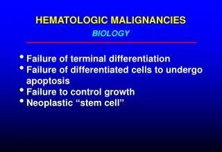
BIOLOGY. HEMATOLOGIC MALIGNANCIES. Failure of terminal differentiation Failure of differentiated cells to undergo apoptosis Failure to control growth Neoplastic “stem cell”. FAILURE OF TERMINAL DIFFERENTIATION. Result: accumulation of rapidly dividing immature cells

HEMATOLOGIC MALIGNANCIES
E N D
Presentation Transcript
BIOLOGY HEMATOLOGIC MALIGNANCIES Failure of terminal differentiation Failure of differentiated cells to undergo apoptosis Failure to control growth Neoplastic “stem cell”
FAILURE OF TERMINAL DIFFERENTIATION Result: accumulation of rapidly dividing immature cells Example: acute leukemias, aggressive lymphomas
FAILURE TO UNDERGO APOPTOSIS Result: accumulation of relatively well-differentiated, slow-growing cells Example: chronic lymphocytic leukemia, indolent lymphomas
THE NEOPLASTIC STEM CELL Propagation of malignant clone may depend on a subset of cells with stem cell-like properties Some neoplastic stem cells retain the ability to differentiate into more than one cell type (eg, myeloproliferative/myelodysplastic disorders) Eradication of neoplastic stem cell essential to cure disease? Neoplastic stem cells may be slow-growing and resistant to treatment
MYELOID NEOPLASIA Myeloproliferative disorders Polycythemia vera Essential thrombocytosis Myelofibrosis/myeloid metaplasia Chronic myelogenous leukemia Myelodysplasia Acute myelogenous leukemia
MYELOPROLIFERATIVE DISORDERS Affected cell: myeloid stem cell All three cell lines affected; clonal hematopoiesis in most cases Differentiation: normal to mildly abnormal Kinetics: effective hematopoiesis Marrow: hypercellular, variably increased reticulin fibrosis Peripheral blood: increase in one or more cell lines in most cases Exception: myelofibrosis
MYELOPROLIFERATIVE DISORDERS Polycythemia Vera Essential Thrombocythemia Myelofibrosis/Myeloid Metaplasia Chronic Myelogenous Leukemia
Polycythemia vera Essential thrombocythemia
MARROW FIBROSIS H&E Reticulin stain
MYELOPROLIFERATIVE DISORDERS Diagnosis usually determined by peripheral blood counts High Hct or platelet count may cause vaso-occlusive symptoms Risk of portal vein thrombosis Splenomegaly, constitutional symptoms frequent Phlebotomy to control high Hct, hydroxyurea or other myelosuppressive Rx to control platelets, constitutional sx, etc Transition to myelofibrosis or acute leukemia possible
Mayo Clin Proc 2004;79:503 SPLENOMEGALY IN MYELOFIBROSIS
JAK2 MUTATION IN CHRONIC MYELOPROLIFERATIVE DISORDERS • Activation of JAK2 tyrosine kinase by cytokines initiates an important signaling pathway in myeloid cells • A single point mutation of JAK2 (Val617Phe) has been identified in a high proportion (65-95%) of patients with polycythemia vera, and also in a substantial proportion of cases of essential thrombocytosis and myelofibrosis • This mutation markedly increases the sensitivity of the cells to the effects of erythropoietin and other cytokine growth factors • Testing for this mutation represents an important diagnostic tool • This finding may lead to development new targeted therapies for myeloproliferative disorders
Diagnostic algorithm for polycythemia vera Mayo Clin Proc 2005;80:947
BIOLOGY CHRONIC MYELOGENOUS LEUKEMIA Virtually all cases have t(9;22) (Ph1 chromosome) or variant translocation involving same genes bcr gene on chromosome 22 fused with abl gene on 9 Fusion gene encodes active tyrosine kinase Clonal expansion of all myeloid cell lines
CHRONIC MYELOGENOUS LEUKEMIA Blood smear Buffy coat Marrow biopsy
LEUKOSTASIS IN CML WBC 300K NEJM 2005;353:1044
Natural history CHRONIC MYELOGENOUS LEUKEMIA Incidence 1:100,000/yr Peak incidence in 40s and 50s Leukocytosis with mixture of mature and immature forms Thrombocytosis common Splenomegaly, constitutional symptoms, eventual leukostasis Transition to acute leukemia (blast crisis) in 20%/yr blasts may be myeloid or lymphoid essentially 100% mortality without BMT
TREATMENT CHRONIC MYELOGENOUS LEUKEMIA Gleevec (imatinib) – inhibits bcr-abl protein kinase Hydroxyurea Alfa interferon Early allogeneic BMT in eligible pts (vs Gleevec Rx?)
MYELODYSPLASIA Affected cell: myeloid stem cell All cell lines affected, clonal hematopoiesis Differentiation: mildly to severely abnormal Morphology and function may be affected Kinetics: Ineffective hematopoiesis (apoptosis of maturing cells in marrow) Marrow: variable cellularity Peripheral blood: decrease in one or more cell lines (usually anemia with or without other cytopenias) Platelets and WBC occasionally increased Cytogenetic abnormalities frequent Risk of transition to acute leukemia high when marrow blast count > 5%
WHO Classification MYELODYSPLASIA Myelodysplastic disorders Refractory anemia Refractory anemia with ringed sideroblasts Refractory cytopenia with multilineage dysplasia Refractory anemia with excess blasts-1 (5-10% blasts) RAEB-2 (10-20% blasts) Mixed myeloproliferative/myelodysplastic disorders Chronic myelomonocytic leukemia Atypical CML (bcr-abl negative)
* SURVIVAL IN MYELODYSPLASIA Overall survival Leukemia-free survival *Mortality of low-risk (RA) patients >70 no different from general population J Clin Oncol 2005;23:7594
Myelodysplasia: marrows showing dyserythropoeisis and hypolobulated megakaryocyte
Myelodysplasia: acquired -thalassemia with Hgb H inclusions in RBC. This is caused by somatic mutations in the -globin gene or an associated regulatory gene, limited to the neoplastic clone Blood 2005;105:443
MDS: micromegakarycyte MDS: hypercellular marrow
RAEB – circulating blast, agranular PMN RAEB – marrow blasts
Myeloblast (red arrow) and abnl RBC precursor (blue arrow) MYELODYSPLASTIC SYNDROME
ACUTE LEUKEMIABiology • Leukemic clone: cells unable to terminally differentiate • May be lymphoid or myeloid • AML: May arise from abnormal stem cell (eg in MDS/MPD) or de novo • Accumulation of immature cells (blasts) • Marrow replaced by leukemic cells • Blasts accumulate in blood and other organs
Pathophysiology ACUTE LEUKEMIA Bone marrow failure fatigue (anemia) infection (neutropenia) bleeding (thrombocytopenia) Tissue infiltration organomegaly skin lesions organ dysfunction pain
Pathophysiology (cont) ACUTE LEUKEMIA Leukostasis (WBC > 50-100K) retinopathy encephalopathy/CNS bleeding pneumonopathy Biochemical effects of leukemic cell products hyperuricemia/tumor lysis syndrome DIC renal tubular dysfunction (lysozymuria) lactic acidosis hypercalcemia (rare) spurious hypoglycemia/hypoxemia/hyperkalemia
Hyperleukocytosis in AML Normal Patient (WBC 250K) NEJM 2003;349:767 26 yo with fever, encephalopathy, retinopathy, dyspnea, lymphadenopathy
Information used in classification ACUTE LEUKEMIA Clinical setting Morphology Histochemistry Surface markers Cytogenetics Molecular genetics
Adverse prognostic features ACUTE LEUKEMIA Old age, poor performance status Therapy-induced Prior myelodysplastic/myeloproliferative disorder High tumor burden Cytogenetics: Ph1 chromosome, deletion of 5 or 7, multiple cytogenetic abnormalities
ACUTE MYELOGENOUS LEUKEMIA Affected cell: myeloid stem cell or committed progenitor cell Differentiation: arrested at early stage, with absent or decreased maturation Kinetics: marrow replacement by immature cells, decreased normal hematopoiesis Marrow: usually markedly hyercellular with preponderance of blast forms Hypocellular variants known Peripheral blood: variable decrease in all cell lines with or without circulating immature cells
Epidemiology ACUTE MYELOGENOUS LEUKEMIA 90% of adult acute leukemia: 2.2 deaths/100,000/yr Incidence rises with age Risk factors: exposure to ionizing radiation, alkylating agents and other mutagens (implicated in10-15% of all cases), certain organic solvents (benzene) Precursor diseases: myelodysplastic & myeloproliferative disorders, myeloma, aplastic anemia, Down syndrome, Klinefelter syndrome, Fanconi syndrome, Bloom syndrome
FAB (French-American-British) classification ACUTE MYELOGENOUS LEUKEMIAS M0 (minimal differentiation) M1 (myeloid blasts) M2 (some differentiation) M3 (promyelocytic) M4 (myelomonocytic) M5 (monocytic) M6 (erythroleukemia) M7 (megakaryoblastic leukemia) Unclassifiable (evolved from MDS, other secondary leukemias) Newer classification schemes place more emphasis on cytogenetics and less on morphology
WHO classification of AML • AML with recurrent cytogenetic abnormalities • t(8;21) • inv(16) • Acute promyelocytic leukemia – t(15;17) and variants • AML with 11q23 (MLL gene) abnormalities • AML with multilineage dysplasia • AML/MDS, therapy-related • AML not otherwise categorized • Minimally differentiated • Without maturation • With maturation • Acute myelomonocytic leukemia • Acute monoblastic and monocytic leukemia • Acute erythroid leukemia • Acute megakaryblastic leukemia • Acute basophilic leukemia • Acute panmyelosis with myelofibrosis • Myeloid sarcoma • AML with ambiguous lineage • Undifferentiated AML • Bilineal AML • Biphenotypic AML
(APML; FAB M3) ACUTE PROMYELOCYTIC LEUKEMIA t (15;17) Translocation involves retinoic acid receptor gene High incidence of DIC/fibrinolysis All-trans retinoic acid induces remission in high proportion of cases Favorable prognosis
M1 M0