Download
1 / 20
210 likes | 984 Views
Discover the intricacies of the sclera - the outer layer of the eye, its layers, diseases like episcleritis & scleritis, and treatment options. Learn about blue sclera discoloration and related syndromes.

E N D
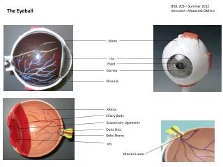
Sclera ** Also known as the white of the eye, is the opaque, fibrous, protective, outer layer of the eye containing collagen and elastic fibers.(it forms 5/6 of the anterior outermost layer of the eye). ** It’s formed from interwoven collagen fibrils of different widths lying within a ground substance and maintained by fibroblasts. ** It’s of variable thickness, 1 mm around the optic nerve head and 0.3 mm just posterior to the muscle insertions.
** In adults, the sclera is white. ** In some children, the sclera is blue due to it’s thinning that shows the pigment cells of the choroid. ** In the elderly, it maybe yellow due to deposition of fat.
Anatomy of the Sclera • The Sclera is divided into 3 layers : 1) The episclera. 2) Scleral stroma. 3) Lamina fusca.
Episclera : • it’s the outermost layer • Anteriorly the episclera consists of a dense, vascular connective tissue which lies between the superficial scleral stroma and Tenon capsule. ** Tenon capsule : the facial sheath that envelopes the eyeball and seperates it from the orbital fat.
2) Scleral stroma : • it’s a dense fibrous tissue with fine elastic fibers. 3) Lamina fusca :it’s the innermost layer of the sclera , it continues with the suprachoroidal and supraciliary lamellae of the uveal tract.
Episcleritis • It’s the inflammation of the superficial layer of the sclera,it’s a common, benign, usually idiopathic, recurrent and frequently bilateral condition. • It causes mild discomfort , and it’s rarely associated with systemic diseases. • It’s usually self-limiting , but as the symptoms are tiresome , topical anti-inflammatory treatment can be given. • In rare severe cases systemic anti-inflammatory drugs maybe given. • And it’s classified into simple and nodular types.
A. Simple Episcleritis • Simple episcleritis accounts for 3/4 of all cases and predominantly affects females. It has a great tendency to recur either in the same eye, or sometimes both together. The attacks become less frequent and after many years disappear completely. ** Presentationis with redness and mild discomfort **SignsRedness may be sectoral or diffuse. Often it has an interpalpebral distribution, in contrast with scleral disease which commonly starts in the upper temporal quadrants. **TreatmentIf mild, no treatment is required.•Cool artificial tears may be adequate in some cases.•A weak topical steroid for 1–2 weeks is usually sufficient.•Oral NSAIDs are sometimes required for 10 days.
B. Nodular Episcleritis • Nodular episcleritis also tends to affect young females but has a less acute onset and a more prolonged course than the simple variety. **Presentation is with a red eye typically first noted on waking. Over the next 2–3 days the area of redness increases in size, becomes more uncomfortable, but remains in the same position. ** Signs one or more tender nodules, almost always within the interpalpebral fissure. **Treatment is similar to that of simple episcleritis.
Simple episcleritisA.sectoral B.diffuse Nodular episcleritis
Scleritis ** Scleritis is an uncommon condition characterized by edema and cellular infiltration of the entire thickness of the sclera. It is much less common than episcleritis and covers a spectrum ranging in severity from self-limiting episodes to a necrotizing disease that may involve adjacent tissues and threaten vision. ** It can be associated with collagen vascular diseases, most commonly rheumatoid arthritis. ** It causes intense occular pain, and both inflammatory and ischemic areas may occur in the sclera.** Scleritis affecting the posterior part of the globe may cause choroidal effusions or simulate a tumor.
** The following may complicate the situation : *Scleral thinning (scleromlalacia), sometimes with perforation.*Keratitis.*Uveitis *Cataract formation.*Glaucoma. ** Treatment may require high doses of systemic steroids or in severe cases cytotoxic therapy and investigation to find any associated systemic diseases.
Infectious scleritis • Infectious scleritis is rare but may be difficult to diagnose because the initial clinical features may be similar to those of immune-mediated disease. In some cases infection may follow surgical or accidental trauma, severe endophthalmitis, or may occur as an extension of primary corneal infection. • Causes : 1)herpes zoster.2)tuberculous scleritis. 3)leprosy.4)syphilis.5)lyme disease.
Blue sclera ** Blue discoloration is caused by thinning or transparency of scleral collagen with visualization of the underlying uvea. **Important causes include the following: • Osteogenesis imperfecta. • Ehlers–Danlos syndrome type VI • Marshall–Smith syndrome , Russell–Silver syndrome , Hallermann–Streiff–François syndrome.
Osteogenesis Imperfecta ** It is an inherited disease of connective tissue, usually caused by defects in the synthesis and structure of Type 1 collagen. There are multiple types, at least two of which have ocular features.
Yellow Sclera ** It manifests in jaundiced patients, it’s not due to pigment on the slcera itself but to accumulation of bilirubin in the vascular cojunctiva.