Introduction to Occlusion
560 likes | 1.33k Views
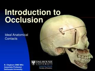
Introduction to Occlusion. Ideal Anatomical Contacts. Which cusp of which tooth is represented by the black dot (red arrow)?. The black dots represent MI contacts in a Class I occlusion. 22 black dots represent 36 areas of contact of mandibular teeth with teeth in the maxillary arch.

Introduction to Occlusion
E N D
Presentation Transcript
Introduction to Occlusion Ideal Anatomical Contacts
Which cusp of which tooth is represented by the black dot (red arrow)? The black dots represent MI contacts in a Class I occlusion 22 black dots represent 36 areas of contact of mandibular teeth with teeth in the maxillary arch
Guidelines for Learning Ideal Contacts Maxillary third molar Maxillary second molar Maxillary first molar Mandibular third molar The DB (distobuccal) cusps of the mandibular molars contact the central pit or fossa of their maxillary counterparts Mandibular second molar Mandibular first molar
Guidelines for Learning Ideal Contacts Maxillary third molar Maxillary second molar Maxillary first molar The MB (mesiobuccal) cusps of the mandibular molars contact the MMR of their maxillary counterparts and the DMR of the tooth mesial to that Mandibular third molar Mandibular second molar Mandibular first molar
Guidelines for Learning Ideal Contacts Maxillary second premolar The B (buccal) cusps of the mandibular premolars contact the MMR of their maxillary counterparts and the DMR of the tooth mesial to that Maxillary first premolar Mandibular second premolar Mandibular first premolar
Guidelines for Learning Ideal Contacts The cusp tip of the mandibular canines contacts the MMR of their maxillary counterparts and the DMR of the tooth mesial to that (maxillary lateral incisor) Maxillary canine Mandibular canine
Guidelines for Learning Ideal Contacts The incisal ridges of the mandibular lateral incisors contact the MMR of their maxillary counterparts and the DMR of the tooth mesial to that (maxillary central incisor) The incisal ridges of the mandibular central incisors contact the MMR of their maxillary counterparts Maxillary lateral incisor Maxillary central incisor Mandibular lateral incisor Mandibular central incisor
Guidelines • Identify tooth shown • Dot represents cusp of opposing tooth • Mandibular teeth are 1/2 cusp mesial to the maxillary counterparts • Contacts same for all mandibular molars • DB cusp (Md) ->Central Fossa (Mx) • MB cusp (Md)-> MMR & DMR (Mx) • B cusps and canines (Md) contact MMR & DMR of teeth 1/2 cusp mesial to maxillary counterparts • Each tooth contacts two teeth • Except maxillary third molars • Except mandibular central incisors
Which cusp of which tooth is represented by the black dot (red arrow)? DB cusp of the mandibular left first molar B cusp of the mandibular right first premolar
Determinants of Occlusion Horizontal Determinants Ridge and Groove Direction
Balancing Working Working
Balancing Working
Balancing Working
Determinants of Occlusion Horizontal Determinants Ridge and Groove Direction Balancing Side Interferences
Balancing Working
Balancing side • Interference • Inner inclines of maxillary MLi cusp (CHC) • Inner inclines of mandibular DB cusp (CHC) Frontal View Balancing Working
Determinants of Occlusion Horizontal Determinants Ridge and Groove Direction Working Side Interferences
Balancing Working
Working side • Interference • Inner inclines of maxillary and mandibular cusps ( Non- CHC cusps) • Outer inclines of maxillary and mandibular cusps (CHC cusps) Frontal View Balancing Working
Determinants of Occlusion Horizontal Determinants Ridge and Groove Direction Distance from the Condyles
Balancing Working
Balancing Working
The angle between the working and idling grooves becomes greater the further the tooth is from the condyles