Download

1 / 48
500 likes | 727 Views
Placenta Previa. Definition. The presence of placental tissue overlying or proximate to the cervical os . Several forms of PP : Complete PP . Partial PP . Marginal PP Low – lying PP . ( within 2-3 cm os . ). Iincidence : PP. 4/1000 pregnancy over 20 weeks Risk factors :

E N D
Definition • The presence of placental tissue overlying or proximate to the cervical os . Several forms of PP : • Complete PP . • Partial PP . • Marginal PP • Low – lying PP . ( within 2-3 cm os . )
Iincidence : PP • 4/1000 pregnancy over 20 weeks Risk factors : _ parity ( 0/2% nullipara – 5% grand multipara _ maternal age : • 0/03 % nullipara 20 < age < 29 • 0/25 % nullipara > 40 year _ number of perior c/s : • ( incidence 10% after 4 or more ) _ number of curettage for spontaneous or induced abortion _ maternal smoking : _ residence at higher altitudes _ male fetus _ multiple gestation ( 39/1000 twin live and 2.8 previa /1000 live) _ gestational age : early pregnancy
Pathogenesis of PP : • Endometrial scarring in the upper segment • Initial tropnoblastic nidation or unidirectional growth into LS . • Increased placental surface to compensate for a reduction in uteroplacental oxygen • the length of lower uterine segment 0/5cm(20 weeks ) , 5 cm ( at term )
Clinical manifestations : • Painless vaginal bleeding ( 70 – 80 % ) VB + uterine contraction : ( 10 – 20 % ) • Asymptomatic (ultrasound ) : ( <10 % ) Initial bleeding : typically 34 weeks • 1/3 : Bleeding prior to 30 weeks Blood transfunsions & preterm delivery & perinatal mortality • 1/3 : VB 30 - 36 weeks • 1/3 : VB after 36 weeks • contraction - vaginal exam - Coitus vaginal Bleeding
Associated conditions : PP malpresentation • PPROM • IUGR : 16% • Congenital anomaly • Placenta Accreta • Velamentous umbilical cord insertion • Vasa previa
Placenta accreta : • 5_ 10 % : with PP • 25 % : PP + one P C/S • 50 % : PP + 2 or more P C/S
Vasa previa : • low lying placenta previa • monochorionic twin gestations • velamentous cord insertion • multi lobed placenta • IVF • Diagnosis : VB + abnormality of FHR (sinusoidal pattern) nucleated RBC • Ultrasound color Doppler vasa previa • Termination : C/S 35- 36 weeks ( corticosteroids )
Velamenous umbilical cord : • Vessels surrounded by fetal memberan,no whartons jelly • 1% singleton • 10% multiple gestation • 25% fetal anomalies • sonography : umbilical cord insertion, 12.5 __ single umbilical artery • Diagnosis : color Doppler , flow • Obstetric complications : IUGR - Prematurity _ congenital anomalies low APGAR scores , fetal bleeding, retained placenta . • Cord compression by fetal descending fetal death . • Pregnancy should not be allowed to proceed beyond 40 weeks .
Diagnosis : PP • Ultrasound • Clinic : Painless VB > 24 weeks
Differential diagnosis : • Third trimester bleeding: 3-4% pregnancy • Abruptio placenta ( 31% ) • PP ( 22% ) • Other cause ( 47% ): labor rupture neoplasm
Ultrasonography Trans vaginal : gold standard _ safe _ effective technique . • accuracy than 99% Trans labial ultrasound • excellent images Trans abdominal ultrasound • accuracy 95% • false negative rate 7% • * an over distended bladder for anterior previa • *for posterior previa : Trendelenburg position
Persistence after second trimester diagnosis : • 10 _ 20 weeks GA 4 _ 6% PP • 10 folds third trimester (0/4 % ) • Amount of overlap • Overlap (20 - 23 w)> 25 mm persistence 40% • Overlap < 14 to 15 mm 20% • Repeat ultrasound: 28 w and 34 w • α-feto Pr: 2 MOM : vaginal bleeding 3rd trimester + preterm labor
MRI : • Posterior previa • High cost • Limited availability
Antepartum management • General principles : • Sonography • Avoidance of coitus & digital cervical examination & exercise & decrease activity • Counseling to seek immediate medical attention if VB
Acute care of symptomatic PP : • admit to the labor • maternal & fetal monitoring • large bore IV & crystalloid & hemodynamic stability & adequate urine out put . • Type BG cross _ match for four units packed blood cells . (Actively bleeding HCT > 30 ) • maternal cardiac monitor: BP &PR every 15 min/h • FHR : continuously monitored . • FHR or FHR or sinusoidal : Anemia & Hypoxia
quantitative monitoring of VB loss • Urine output : hourly with Foley catheter • Laboratory monitoring • HB-HCT /q 4 -6 h • Serum electrocytes & indexs of renal function:every 6-8 / h • PT _ PTT _ CBC _ PLT- fibrinogen
Unstable hemodynamic or underlying disease (cardiac& pulmonary) place swan Ganz catheter ( CVP ) • ( PCWP ) & cardiac out put • Tocolysis is not administeral to VB If : VB or ceased • Rh D imminuglobolin
Conservation management of stable preterm patients • Hospitalized at bed rest • minimize constipation ( high fiber diet & stool softens ) • Ferrous gluconate supplements ( 3- 4 time/day ) + vitamin C • Periodic maternal HCT • Maternal blood sample type, cross match ( 2 _ 4 units P.C ) HCT > 30 • Corticosteroid therapy : ( 24 _ 34 weeks ) • RH ( D ) _ immunoglobolin : ( 3 weeks ) • Fetal Heart rate monitoring : • sonography : IUGR _ AF _ placenta location • Tocolysis : contraction ( Mg so4 4 H2o ) • Cervical cercelage : longer gestations, heavier birth weight, , reduction in antenatal hospitalization .
PPROM & PP : • Tocolysis : controversial _ hemodynamically stable & uninfected women • Corticosteroid < 32 weeks
Out patient management : • 48 h after stopped bleeding • Restriction activity • Live within 15 min of the hospital • Have an adult companion available 24h/day ( for transport & call ambulance ) • Be reliable & able to maintain bed rest at home . • understand the risks of PP . • Benefits of out patients • Longer duration of pregnancy ( 33- 36 w ) higher mean birth weight • Lower over all cost
Delivery Timing : • FHR • Life threatening material hemorrhage • After 34 weeks : presence fetal pulmonary maturity . (bleeding) • Amniocentesis at 36 weeks : repeat every week .
Procedure : • Abdominal delivery ( choice) • NVD : fetal demise _ previable fetus hemodynamically stability , low lying previa . • Available 2 to 4 units PC . • Method of CS : • Surgical instruments : CS hysterectomy • 5 - 10 % risk placenta accreta . • Pre operative sonographyic localization of placenta . • Incised placenta : delivered rapidly & cord clamped to hemorrhage from fetus .
Anesthesia : GA for emergency CS • Regional A for stable patients • RH ( D ) negative women • RH ( D ) _ Immune globulin .
Out come PP : • General • Maternal mortality : 1 % • perinatal mortality : 10 % • Principal causes of prenatal mortality • * Preterm delivery • *Fetal anemia • *Hypoxia • *Growth restriction • Recurrence rate :4 _ 8 %
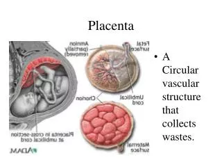
Introduction : • A.P : premature separation of a normally implanted placenta after 20 weeks but prior to delivery infant . Immediate cause : • Rupture of maternal vessels in decidua • basallis Rare cause : • Bleeding fetal _ placenta vessels . • Separation of placenta : hematoma • Retro placenta complete or partial exchange gases nutrient to the fetus
Incidence • 0/4 to 1/3% ( 1/75 _ 1/225 ) • Sever AP to still birth : 1/ 830 • Prenatal mortality 25 folds • Neurological sequel 15% • CP 20% ( non AP 1%) • 1/3 antepartum bleeding ___ AP • Pathogenesis : • Catastrophic trauma • PPROM • Chronic pathologic vascular process ( IUGR _ preterm labor )
Risk factors : mechanical factors : Truma : external compression decompression , rapid acceleration _ deceleration present within 24h of event Monitoring : 4_ 6 h period ( VB _ tenderness ) Fetal maternal transfusion Sudden internal decompression of the uterus : PPROM Placental implantation over uterine anomaly or myoma Hypertension : PIH & chronic, 5 folds sever Abruption • Antihypertensive therapy dose not reduce risk of Abruption • Mg So4 AP
Cigarette smoking : 2.5 fold sever A.P Risk : 40% / pocket / day Mechanism : ischemic peripheral necrosis of decidua cigarette smoker & hypertension are synergistic . Maternal age & parity 2.5 % • Endometrial scarring & impaired decidualization Cocaine abuse : 10% • Acute vasoconstriction ischemia reflex vasodilation bleeding
PPROM : 2 - 5 % AP • Infection or oligohydramnios 7 to 9 fold AP • Abruption thrombin proteas PPROM
Inherited thrombophilia : 1/5 – 12 folds • factor V leiden: • maternal venous thromboembolism , fetal death ,IUGR , sever PIH , abruption • Prc ,Prs , Antithrombin • VII , VIII , IX , XI • Hyperhomocysteinemia : 31% Ab • Congenital hypofibrinogenemia afibrinogenemia, XIII AP
Previous Abruption : • Ten folds . AP Multifetal gestation & polyhydramnios • 3 folds AP • cause : rapid uterine decompression upon delivery of one twin . Others : • folate deficiency , leiomyoma , circumvallata placenta
Clinical manifestation • VB > 80% • Abdominal pain > 50% • Uterine contraction • Uterine tenderness 66% • FHR 60% • Uterine tone 17% • Back pain : posterior placenta 60% • Preterm birth 22% • Chronic abruption • IUFD 15% • Shock
Concealed hemorrhage • 20% • placental margins remain adherent • The fetal membrane retain their attachment to the uterine wall • The fetal head obstruct cervical os
Coagulopathy • sever abruption with death fetus 20% coagulopathy • hypofibringenemia < 150mg/dl • D- Dimer , FDP > 100 ng/ml, PLT • Kidney : ATN
Diagnosis : • Clinic • Sonography _ difficult : 25% • Laboratory not useful _ CA 125, D- Dimer _ thrombo modolin -Fibrinogen -PLT • Pathologic findings: • Clot depression Maternal surface of placenta
Differential diagnosis : • Placenta previa • Vasa previa • Labor • Uterine rupture • Cervicovaginal neoplasm • Abdominal disorder ( pain without bleeding )
Management • Initial approach : • Closely monitoring • Large _ Bore IV • Maternal hemodynamic status: • BP- PR-Out Put - BG Rh- HCT- PLT-Fib- PT- PTT • Normotensive + normal HCT & Abruption : • Previousely hypertensive & acute bleeding
management • Fetal monitoring • Crystalloid infusion • RBC , packed cells • 300 cc packed cell 200 cc RBC 3-4% HCT • PT & PTT( 1/5 times): 2 units FFP • 5 units packed cell: PTT- PT - fibrinogen - PLT • PLT < 50,000 : 6 units of PLT • Tocolysis: contraindication ( sever abruption, DIC)
Delivery : optimal treatment • Mild Abruption+ preterm : Expectant management • Corticosteroid therapy < 34 weeks • tocolysis < 34 weeks
Labor : • Monitoring on labor room . • Mode & timing delivery : • Condition & gestational age • Condition ( BP , DIC , Hemorrhage status of cervix , FHR ) • VD : Amniotomy _ Internal monitoring of fetus & intrauterine pressure catheter • Pressure > 25 mmHg abnormal uterine flow oxygenation of fetus • Poor condition sever hypertone , hemorrhage ,DIC, FHR • C/S : HCT > 30% , fibrinogen (150- 200 mg/dl ), PLT > 60,000 • Anesthesia : GA • Appropriate mode of delivery : C/S • ( VD : cervical dilatation in Parous women)
Out come • Perinatal mortality 20% (still birth, 50% placenta separation) • IUGR • Prematurity : 4 folds • C/S : 3 /4 delivery ( Sweden ) • Midtrimester abruption poor prognosis • Recurrence risk : 5 _ 15 % • Base line risk : o/4% to 1/3% • Two abruption: risk 25% • Sever abruption: ( dead fetus ) 7% • Abruption & subsequent pregnancy : • Abruption • SGA • Preterm labor • PIH