Download

1 / 25
340 likes | 775 Views
PLACENTA PREVIA. DEFINITION. When the placenta is implanted partially or completely over the lower uterine segment(over and adjacent to the internal os)it is called placenta previa. INCIDENCE. O.5 – 1% among hospital deliveries 80% cases found in multiparous women

E N D
DEFINITION When the placenta is implanted partially or completely over the lower uterine segment(over and adjacent to the internal os)it is called placenta previa
INCIDENCE • O.5 – 1% among hospital deliveries • 80% cases found in multiparous women • Increase incidence beyond 35yrs • Increase incidence with high birth order and multiple pregnancy
ETIOLOGY THEORIES POSTULATED • Dropping down theory • Persistence of chorionic activity • Defective decidua • Big surface area of the placenta
ETIOLOGY…..Contd HIGH RISK FACTORS • Multiparity • Increased maternal age • Previous cesarean section or any other scar in the uterus • Placental size and abnormality • Smoking • Prior curettage
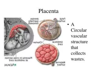
PATHOLOGICAL ANATOMY • PLACENTA • Large and thin • Tongue shaped extension from the main placental mass • Extensive area of degeneration and calcification • Placenta may be morbidly adherent • UMBILICAL CORD • Attached to the margin or into the membrane • Insertion of the cord may be close to the internal os • Fetal vessels may run across the internal os • LOWER UTERINE SEGMENT • Increased vascularity • The lower uterine segment and the cervix becomes soft and more friable
TYPES OR DEGREE • TYPE I – Low – lying • Major part of the placenta is attached to the upper segment • Only the lower margin encroaches to the lower segment • But not up to the os • TYPE II – Marginal • Placenta reaches the margin of the internal os • But does not cover it • TYPE III – Incomplete or partial central • Placenta covers the internal os partially • TYPE IV – Central or total • Placenta covers the internal os even after it is fully dilated
CLINICAL FEATURES SYMPTOMS • VAGINAL BLEEDING • Sudden onset • Painless • Causeless • Recurrent SIGNS • General condition and anemia are proportionate to the visible blood loss
CLINICAL FEATURES……Contd ABDOMINAL EXAMINATION • The size of the uterus proportionate to the period of gestation • The uterus feels relaxed, soft and elastic without any localised area of tenderness • Persistence of malpresentation • Head is floating • Fetal heart sound • Stallworthy’s sign
CLINICAL FEATURES……Contd VULVAL INSPECTION • Bright red or dark coloured • Amount of blood loss VAGINAL EXAMINATION MUST NOT BE DONE OUTSIDE THE OT
CONFIRMATION OF DIAGNOSIS • LOCALISATION OF PLACENTA • SONOGRAPHY • TAS • TVS • Transperineal ultrasound • Color Doppler flow study • MAGNETIC RESONANCE IMAGING • CLINICAL • By internal examination(double set up examination) • Direct visualization during cesarean section • Examination of the placenta following vaginal delivery
COMPLICATION MATERNAL • DURING PREGNANCY • APH • Malpresentation • Premature labor • DURING LABOUR • Early rupture of the membrane • Cord prolapse • Slow dilation • Intrapartum haemorrhage • Increased incidence of operative interference • PPH • PUERPERINM • Sepsis • Subinvolution • Embolism
COMPLICATION FETAL • Low birth weight • Asphyxia • Intrauterine death • Birth injuries • Congenital malformation
MANAGEMENT • PREVENTION • Adequate antenatal care • Antenatal diagnosis • Warning haemorrhage should not be ignored • Colour doppler USG
MANAGEMENT…..Contd • AT HOME • Put to bed • To assess the blood loss • Quick but gentle abdominal examination • Vaginal examination must not be done • TRANSFER TO HOSPITAL • Emergency • Dextrose saline drip • Accompanied by persons for donation • ADMISSION TO HOSPITAL • Considered as APH
MANAGEMENT…..Contd • IMMEDIATE ATTENSION • Amount of the blood loss • Blood samples are taken • A large bore IV cannula is sited • Infusion of NS • Gentle abdominal palpation • Inspection of vulva
MANAGEMENT…..Contd • FORMULATION OF LINE OF TREATMENT • Expectant management • Active management
EXPECTANT MANAGEMENT • VITAL PREREQUISITES • Availability of blood transfusion • Facilities for cesarean section throughout 24hrs • SELECTION OF CASES • Mother is in good health status • Duration of pregnancy less than 37 weeks • Active vaginal bleeding is absent • Fetal well being is assured
EXPECTANT MANAGEMENT…..Contd • CONDUCT OF EXPECTANT TREATMENT • Bed rest with bathroom privileges • Investigations • Periodic inspection • Supplementary hematinics • A gentle speculum examination • Use of tocolysis • Rh immunoglobulin • Termination done at 37 weeks • Steroid therapy
ACTIVE MANAGEMENT • INDICATIONS • Bleeding occurs at or after 37 weeks of pregnancy • Patient is in labour • Patient is exsaguinated state on admission • Bleeding is continuing and of moderate degree • Baby is dead or known to be congenitally malformed
DEFINITIVE MANAGEMENT • CESAREAN DELIVERY • Placental edge is within 2cm from the internal os • VAGINAL DELIVERY • Placental edge is clearly 2-3cm away from the internal os