Download
1 / 54
580 likes | 681 Views
This study delves into the intricate process of cardiovascular system development, from the establishment of the primordial network to the morphogenesis of the heart chambers. It explores the formation of the endothelial tube meshwork, the development of the cardiac tubes, and the partitioning of heart chambers, shedding light on the blood circulation of the fetus and congenital cardiovascular defects. Step-by-step, it unravels the journey from the endothelial tube network fusion to the formation of the primitive heart and the growth of the bulbus cordis, ventricle, and atrium.

E N D
Development of the Cardiovascular System
Contents • Establishment of the primordial cardio-vascular system • Development of the heart • Blood circulation of fetus and circulatory changes after birth • Congenital defects of the cardiovascular system
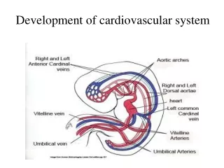
Endothelial tube meshwork • Yolk sac • mesenchyme cells blood islands Central C Peripheral C Primitive Blood cell Endothelial C Blood C Endothelial tubule
Endothelial tube network appears in chorion and body stalk, and connect to vitelline circulation. • By the 18-20th days, endothelial tube network appears in intraembry-onic mesenchyma to form intraembryonic endothe-lial tube network.
By the end of 3rd week, intraembryonic and extra-embryonic endothelial tube networks connect to each other. • Endothelial tube networks fuse or disappear to form primordial cardiovas-cular system.
Primitive heart cardiac tubes 4w End of 4w 20d • ① cardiac tube:2 tubes1 tubeprimitive heart • ② arteries • ③ veins
Dorsal A Dorsal A Aortic arches Dorsal aorta 20d 4w End of 4w Umbilical A Vitelline A • ① cardiac tube • ② arteries • ③ veins 2 dorsal A 1 ,many branches Few pairs of vitelline A 1 pair of umbilical A 6 pairs of aortic arches
Anterior cardinal V Posterior cardinal V Vitelline V Common cardinal V Umbilical V 20d 4w End of 4w • ① cardiac tube • ② arteries • ③ veins 1 pair of anterior cardinal V 1 pair of posterior cardinal V 1 pair of vitelline V 1 pair of umbilical V Common cardinal V
Development of the Heart • Development of the cardiac tube • Morphogenesis of the heart • Partitioning of heart chambers
Cardiogenic area Oropharyngeal membrane • Development of the cardiac tube • Cardiogenic areais anterior to the oropharyngeal membrane.
Pericardial cavity cardiaogenic plate 18~19th day • A cavity appears in the cardiogenicarea • --pericardial cavity • B. Ventral of the cavity is cardiaogenic cords • --cardiaogenic plate
Pericardial cavity cardiac tube 20th day • C. cardiaogenic plate becomes hollow --cardiac tube
cardiac tube Pericardial cavity 22nd day D. Cephalic folding: Pericardial cavity: dorsal → ventral Cardiac tube: ventral → dorsal
E. Lateral folding: 2 cardiac tubes → single cardiactube. F. The tube remains attached to the dorsal side of the pericardial cavity by the dorsal mesocardium.
Caudal end Cephalic end cardiac tube Pericardial cavity Transverse sinus G. The dorsal mesocardium breaks down to form the transverse sinus, which connects both sides of the pericardial cavity. Cephalic end Arteries,Caudal end Veins
Artery end Cardiac tube Vein end The 21st d • Morphogenesis of the heart • Part of the cardiac tubes merged • Cephalic end A • Caudal end V
bulbus cordis ventricle atrium • Cardiac tubes almost merged • Three expansions • Bulbus cordis • Ventricle • Atrium The 22nd d
truncus arteriosus bulbus cordis ventricle atrium sinus venosus The 23rd d • The 4th expansion, the sinus venosus appears • The truncus arteriosus appears • The bulbus cordis and ventricle grow faster than other regions,the cardiac tube starts to bend.
truncus arteriosus sinus venosus The 24th d • Form a ‘U’ like structure, the cardiac loop--bulboventricular loop.
Aortic arches atrium ventricle The 35th d • The bulboventricular loop continues to grow and bend: • Atrium shifts in dorso-cranial direction and bulges laterally on each side of bulbus. • Sinus venousus located at caudal portion of atrium
Aortic arches atrium ventricle The 35th d • Primary ventricle develop into the left ventricle. • The bulbus cordis proximal portion develops into the right ventricle. • Atrioventricular canal: atrioventricular junction remains narrow. The normal heart shape was established, but partitioning has not completed.
Partitioning of Heart Chambers (from 27th day to 37th day) • Partitioning of atrioventricular canal • Partitioning of the primitive atrium • Partitioning of the primitive ventricle • Division of truncus arteriosus and bulbus cordis
Endocardiac cushion: • The endocardial cushions grow toward each other and fuse • Partitioning of atrioventricular canal
Endocardiac cushion Bicuspid Ttricuspid • Lateral atrioventricular cushion: form atrioventricular • valve. Left → bicuspid, right →tricuspid
Septum primum Foramen primum Endocardiac cushion • Partitioning of the primitive atrium • Septum primum: a thin sickle-shaped crest appearing from dorso-cranial wall of atrium. • Foramen primum: septum primum grows toward the endocardial cushions, leaving an opening between its lower edge and the endocardial cushions End ofthe 4th w
Small holes Foramen primum Early 5th w
Septum secundum Foramen secundum Septum primum End of the 5th w • Foramen secundumSmall holes fuse to form the foramen se-cundum,The foramen primum closed. • Septum secundum: another membraneappears on the right of the septum primum.
Septum secundum Foramen secundum Septum primum Foramen ovale • Foramen ovale : septum secundum extends downward to cover the foramen secundum, but leaving an opening. • The septum primum covers the foramen ovale, serves as a valve. Early 6th w
Before birth, blood can flow from right atrium toward the left atrium • After birth, two septums fuse , the foramen ovale closed complete, and atrium is separated into R and L atria.
Partitioning of the primitive ventricle • The muscularInterventricular septum grows up from the floorof the ventricle. EC LV RV Inter-ventricular septum The 4th w
Interventricular foramen • the muscular interven- • tricular septum keeps • growing toward endo- • cardial cushions, but left • an opening, called inter- • ventricular foramen. EC Inter- ventricular foramen IV septum End of the 5th w
Membranous interventricular septum: Derived from right bulbar ridge, left bulbar ridge and the endocardial cushion,closes the interventricular foramen Membranous interventricular septum endocardial cushions End of the 7th w
The interventricular septum: muscular partion + membranous portion Left ventricle Pulmonary artery Right ventricle Aorta
Truncal ridge Truncus arteriosus Bulbar ridge Bulbuscordis • Division of truncus arteriosus and bulbus cordis The 5th w • Two spiral truncal ridges/ bulbar ridges grow from the inner walls of the truncus arteriosus and bulbus cordis.
Aorta Pulmonary artery Aortico- pulmonary septum • These ridges twist around each other and fuse to form a spiral aorticopulmonary septum.
The ridges spiral neatly down the truncus until they reach the ventricles.
As the same time, the division of the ventricle is completed. • Aorticopulmonary septum divides truncus arteriosus and bulbus cordis into two channels: pulmonary trunkconnecting to the right ventricle;aortaconnecting to the left ventricle.
Blood circulation of fetus and circulatory changes after birth
Before birth Placental circulation: umbilical A. & V.
Circulatory changes after birth a.Umbilical A: distal parts becomes into medial umbilical ligament, but proximal portions persist as superior vesical arteries. b. Umbilical V and ducts venousus: constrict and becomes into ligamen-tum teres hepatis and ligamentum venosus c. Ductus arteriosus: constrict and become ligamentumarteriosum d. Foramen ovale closed
Atrial septal defect • An atrial septal defect (ASD) is a common congenital heart anomaly. • The most common form of ASD is patent oval foramen. • Consequently there is a mixing of oxygenated and deoxygenated blood.
Atrial septal defect • Perforation of valve of ovale foramen • Excessive resorption of the septum primum • Inadequate development of the septum secundum. • B+C • Endocardial cushion defect with septum primum defect
Ventricular septal defect • Muscular part of the ventricular septum: sparsely • Membranous part of the ventricular septum defect:commonly Unfused endocardial cushion,bulbar ridge and muscular septum or over absorption of membranous septum
Patent ductus arteriosus • Ductus arteriosus fails to be closed after birth. • Isolated or combined with other defects. • Blood will be shunted from the aorta to the lungs, eventually causing irreversible pulmonary hypertension.
Persistent truncus arteriosus • Aorticopulmonary septal ridges fail to fuse and descend; • Truncus overrides both ventricles; • Accompanied by ventricular septal defect; • Resulting in cyanotic defect.