Slide Note
0 likes | 91 Views
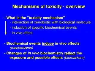
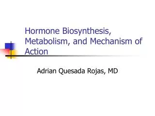
Adrenal cortex secretes corticosteroids with glucocorticoid, mineralocorticoid, and weakly androgenic activities. They are synthesized from cholesterol, regulating fluid-electrolyte balance, carbohydrate metabolism, and stress response. Glucocorticoids like cortisol impact glucose, protein, and fat metabolism, while mineralocorticoids like aldosterone maintain Na+ and fluid balance. Glucocorticoids also have anti-inflammatory, stress-resistant, and immunosuppressant effects. Pharmacokinetics involve oral absorption, metabolism, and excretion. They are used in replacement therapy for adrenal insufficiency and pharmacotherapy for conditions like arthritis, collagen diseases, and severe allergic reactions.

E N D
CORTICOSTEROIDS SUHAS AGEY ASSISTANT PROFESSOR DEPARTMENT OF PHARMACOLOGY HRPIPER, SHIRPUR
CONTENTS Introduction Biosynthesis Classification Mineralocorticoids Glucocorticoids Mechanism of action Androgens
INTRODUCTION The adrenal cortex secretes steroidal hormones which have glucocorticoid, mineralocorticoid and weakly androgenic activities. The corticoids (both gluco and mineralo) are 21 carbon compounds having a cyclopentanoperhydro-phenanthrene (steroid) nucleus. They are synthesized in the adrenal cortical cells from precursor molecule -cholesterol. Adrenal steroidogenesis takes place under the influence of ACTH which makes more cholesterol available for conversion to pregnenolone and induces steroidogenic enzymes.
CORTICOSTEROIDS They maintain fluid-electrolyte. cardiovascular and energy substrate homeostasis as well as functional status of skeletal muscles and nervous system. They prepare the body to withstand effects of all kinds of noxious stimuli and stress. Glucocorticoid Effects on carbohydrate, protein and fat metabolism. Mineralocorticoid Effects on Na+ K+ and fluid balance.
A. MINERALOCORTICOIDS The principal mineralocorticoid (aldosterone) action is enhancement of Na+ and water reabsorption from the distal convoluted tubule in kidney. There is an associated increase in K+ and H+ excretion. Its deficiency results in decreased maximal tubular reabsorptive capacity for Na+; kidney is not able to retain a even in the Na+ deficient state so that Na+ is progressively lost.
B. GLUCOCORTICOIDS As the name implies, glucocorticoids regulate the glucose homeostasis Glucocorticoids consist of 3 hormones 1. Cortisol (also called hydrocortisone) 2. Corticosterone and 3. Cortisone Glucocorticoids are usually released in response to stressful stimuli, hence considered as a stress-relieving hormone.
ACTION OF GLUCOCORTICOIDS 1. Glucose formation- Glucocorticoids promote glycogen deposition in liver by promoting gluconeogenesis. They inhibit glucose utilization by peripheral tissues. results in hyperglycaemia and a diabetes-like state. 2. Protein breakdown. They cause protein breakdown and amino acid mobilization from peripheral tissues. These AA used up in gluconeogenesis and excess urea is produced resulting in negative nitrogen balance. 3. Lipolysis. Glucocorticoids stimulate lipolysis, the breakdown of triglycerides and release of fatty acids from adipose tissue into the blood.
ACTION OF GLUCOCORTICOIDS Anti-inflammatory effects. Glucocorticoids inhibit white blood cells that participate in inflammatory responses. Unfortunately, glucocorticoids also retard tissue repair; as a result, they slows wound healing. 5. Resistance to stress. The additional glucose supplied by the liver cells provides tissues with a ready source of ATP to combat a range of stresses, including exercise, fasting, fright, temperature extremes, high altitude, bleeding, infection, surgery, trauma, and disease. 6. Immunosuppressant response. High doses of glucocorticoids depress immune responses. For this reason, glucocorticoids are prescribed for organ transplant recipients to retard tissue rejection by the immune system 7.
Corticosteroids mechanism of action The aldosterone penetrates the cell membrane and binds to the Gluco/mineralocorticoid receptor (G/M-R) The aldosterone binds to the steroid binding domain near the carboxy terminus. Binding of the steroid to G/MR dissociates the complexed proteins (HSP90) removing their inhibitory influence on it. The steroid bound receptor dimer translocates to the nucleus, and interacts with specific DNA sequences called ‘mineralocorticoid responsive elements' (MREs) within the regulatory region of appropriate genes. GC-regulation of protein synthesis MC- directs synthesis of proteins (aldosterone-induced proteins-AIP-Na+K+ATPase).
PHARMACOKINETICS All corticoids, except DOCA are absorbed and are effective by the oral route. Hydrocortisone undergoes high first pass metabolism, has low oral: parenteral activity ratio. Oral bioavailability of synthetic corticoids is high. Hydrocortisone is 90% bound to plasma protein, mostly to a specific cortisol-binding globulin The corticosteroids are metabolized primarily by oxidation followed by conjugation with glucuronic acid or sulfate and are excreted in urine.
USES A. Replacement therapy 1. Acute adrenal insufficiency It is an emergency. Hydrocortisone (100 mg) /dexamethasone (8-16 mg) are given i.v. 2. Chronic adrenal insufficiency (Addison's disease) Hydrocortisone given orally is the most commonly used drug. B. Pharmacotherapy 1. Arthritis (i) Rheumatoid arthritis, (ii) Osteoarthritis, (iii) Rheumatic fever, (iv) Gout etc 2. Collagen diseases- Systemic lupus erythematous (SLE), dermatomyositis, nephritis, glomerulonephritis etc. 3. Severe allergic reactions Corticoids used in anaphylaxis, angioneurotic edema, urticaria and serum sickness. 4. Autoimmune diseases Autoimmune haemolytic anaemia, thrombocytopenia , chronic hepatitis Prednisolone 1- 2 mg/kg/day. 5. Bronchial asthma- glucocorticoid is given Cases of Status asthmaticus, Acute asthma exacerbation, Severe chronic asthma; i.v.
6. Infective diseases- They are indicated in conditions like severe forms of tuberculosis, severe lepra reaction, bacterial meningitis and pneumonia inAIDS patients. 7. Eye diseases- allergic conjunctivitis, iritis, iridocyclitis, keratitis etc; Topical instillation given as eye drops 8. Skin diseases- dermatitis, Stevens-Johnson syndrome and other severe afflictions. 9. Intestinal diseases- Ulcerative colitis, Crohn 's disease, coeliac disease are inflammatory bowel diseases. 10. Neurological conditions Cerebral edema due to tumours, tubercular meningitis, etc., responds to corticoids. 11. Malignancies Corticoids are an essential component of combined chemotherapy of acute lymphatic leukaemia, Hodgkin 's and other lymphomas 12. Organ transplantation- High dose corticoids are given along with other immuno-suppressants to prevent the rejection reaction. 13. Septic shock- low-dose (hydrocortisone 100 mg 8 hourly i.v. infusion for 5-7 days)
ADVERSE EFFECT A. Mineralocorticoid- Sodium and water retention, edema, hypokalaemic alkalosis and a progressive rise in BP. B. Glucocorticoid 1. Cushing's syndrome: 7. Osteoporosis: 2. Hyperglycaemia 8. Glaucoma: 3. Muscular weakness: 9. Growth retardation: 4. Susceptibility to infection: 10. Foetal abnonnalities: Glucocortlcoid antiprogestin mefepristone, also acts as a glucocorticoid receptor antagonist. In Cushings syndrome, it can suppress the overdose of corticosteroid. antagonist The 5. Delayed wound healing 11. Psychiatric disturbances: 6. Peptic ulceration
INTRODUCTION- GLANDS EXOCRINE GLANDS Exocrine glands are glands that secrete their products into ducts. EXAMPLE: Sweat glands Salivary glands Mammary glands Stomach Liver etc.
INTRODUCTION- GLANDS ENDOCRINE GLANDS- Glands that secrete hormones directly into the blood circulation and not through a duct. EXAMPLE: Pituitary gland Thyroid gland Parathyroid Gland Adrenal glands Pancreas Pineal gland Thymus Gland Gonads (Testes/ Ovaries)
HORMONES A hormone is a molecule that is released in one part of the body but regulates the activity of cells in other parts of the body by entering into the blood circulation. FUNCTIONS Help maintain homeostasis, despite imbalance, such as infection, trauma, emotional stress, dehydration, starvation, hemorrhage, and temperature extremes. Help regulate metabolism and energy balance. Help regulate contraction of smooth and cardiac muscle and secretion by glands. Play a role in the smooth, sequential growth and development of the human body. Contribute to the basic processes of reproduction, including gamete production, fertilization, nourishment of the embryo and fetus, delivery, and nourishment of the newborn.
CHEMICAL CLASSIFICATION OF HORMONES Water Soluble Lipid Soluble Amine Hormones- A. Catecholamines Epinephrine, Norepinephrine and dopamine B. Other- Melatonin, Histamine Steroid Hormones- Estrogen, Progesterone, Testosterone, Aldosterone, corticoids Thyroid Hormones- T3 and T4 Protein or Peptide Hormones- Growth Hormones, Insulin, Oxytocin, ADH, TSH, ACTH, FSH, LH, Prolactin, PTH, Calcitonin The Gas Nitric Oxide (NO)
MECHANISMS OF HORMONE ACTION –A. LIPID SOLUBLE HORMONES Lipid soluble Hormones - includes steroid hormones, bind to receptors within target cells. A free lipid-soluble hormone molecule diffuses from the blood, through interstitial fluid, and through the lipid bilayer of the plasma membrane into a cell. the hormone binds to and activates receptors located within the cytosol or nucleus. The activated receptor– hormone complex then alters gene expression: It turns specific genes of the nuclear DNA on or off. The new proteins alter the cell’s activity and cause the responses typical of that hormone
MECHANISMS OF HORMONE ACTION- B. WATER SOLUBLE HORMONES Water-soluble hormones (amine, peptide or protein) bind to receptors that present on the cell surface/ plasma membrane When a water-soluble hormone binds to its receptor at the outer surface of the plasma membrane, intracellular changes takes place. These changes leads to series of activation or inactivation of intracellular enzymes or proteins or ion channels (Signal transductions) It results into the physiological response.
PITUITARY GLAND The pituitary gland or hypophysis is a pea-shaped structure that lies in the hypophyseal fossa of the sphenoid bone attached to the hypothalamus by a stalk called the infundibulum. It is also called the “Master” endocrine gland because it secretes several hormones that control other endocrine glands.
PITUITARY GLAND Anatomically and functionally, the pituitary gland is divided into 2 parts Anterior pituitary (Adenohypophysis) secretes hormones that regulate a wide range of bodily activities and are stimulated by releasing hormones and suppressed by inhibiting hormones from the hypothalamus. Posterior pituitary (Neurohypophysis): - It is consist of neurons that store and release two hormones.
PITUITARY HORMONES Anterior lobe: Secretary cells & their Hormones Posterior lobe: Vasopressin (ADH) oxytocin Somatotrophs - Growth hormone (GH) & Insulin-like Growth Factor (IGF) Thyrotrophs- Thyroid-stimulating hormone (TSH) Corticotrophs-Adrenocorticotrophic hormone (ACTH) Lactotrophs- Prolactin Gonadotrophs- Follicle-stimulating hormone (FSH) & Luteinizing hormone (LH)
HYPOTHALAMUS AND PITUITARY GLAND Hypophyseal Portal System The neurosecretory cells in the hypothalamus synthesize the releasing and inhibiting hormones in their cell bodies. Hypothalamic hormones that release or inhibit anterior pituitary hormones reach the anterior pituitary through a portal system. The superior hypophyseal arteries, branches of carotid arteries, bring blood into the hypothalamus. These arteries divide into a capillary network called the primary plexus. From the primary plexus, blood drains into the hypophyseal portal veins that pass down the anterior pituitary, the hypophyseal portal veins divide again and form another capillary network called the secondary plexus.
ANTERIOR PITUITARY- HUMAN GROWTH HORMONE AND INSULIN-LIKE GROWTH FACTORS (IGF) The main function of hGH is to promote the synthesis and secretion of small protein hormones called insulin-like growth factors (IGF) in the liver, skeletal muscles, cartilage, bones etc. It stimulates the growth and division of most body cells but especially those in the bones and skeletal muscles. Body growth in response to the secretion of GH is evident during childhood and adolescence, and thereafter secretion of GH maintains the mass of bones and skeletal muscles.
ANTERIOR PITUITARY- A. HUMAN GROWTH HORMONE AND INSULIN-LIKE GROWTH FACTORS (IGF) Protein Metabolism- hGH and IGF is a protein anabolic hormone. IGFs cause cells to grow and multiply by increasing uptake of amino acids into cells and accelerating protein synthesis. IGFs also decrease the breakdown of proteins and the use of amino acids forATP production. Carbohydrate metabolism- IGFs and hGH may also stimulate liver cells to release glucose into the blood. Lipid Metabolism- IGFs and hGH enhance lipolysis in adipose tissue, which results in increased use of the released fatty acids forATP production by body cells.
CONTROL OF GROWTH HORMONES Somatotrophs in the anterior pituitary release bursts of GH every few hours, especially during sleep. Their secretary activity is controlled mainly by two hypothalamic hormones: (1)growth (GHRH) promotes secretion of human GH, & (2) growth hormone–inhibiting (GHIH) suppresses it. hormone–releasing hormone hormone A major regulator of GHRH and GHIH secretion is the blood glucose level
ANTERIOR PITUITARY- B. THYROID STIMULATING HORMONE (TSH) Thyroid-stimulating hormone (TSH) stimulates the synthesis and secretion of the two thyroid hormones, triiodothyronine (T3) and thyroxine (T4), both produced by the thyroid gland. Thyrotropin-releasing hormone (TRH) from the hypothalamus controls TSH secretion. The release of TRH in turn depends on blood levels of T3 and T4; high levels of T3 and T4 inhibit secretion of TRH via negative feedback.
ANTERIOR PITUITARY- C. ADRENOCORTICOTROPIC HORMONE (ACTH) Corticotropes secreteACTH. ACTH controls the production and secretion of glucocorticoids (cortisol) by the cortex of the adrenal glands. Corticotropin-releasing hormone (CRH) from the hypothalamus stimulates the secretion of ACTH by corticotropes. Stress-related stimuli, such as low blood glucose or physical trauma. Glucocorticoids inhibit CRH and ACTH release via negative feedback
ANTERIOR PITUITARY- D. FOLLICLE-STIMULATING HORMONE (FSH) AND LUTEINIZING HORMONE (LH) i. Follicle-Stimulating Hormone In females, the ovaries are the targets for follicle-stimulating hormone (FSH). Each month FSH initiates the development of several ovarian follicles. In males, FSH stimulates sperm production in the testes. Gonadotropin-releasing hormone (GnRH) from the hypothalamus stimulates FSH release. The release of GnRH and FSH is suppressed by estrogens in females and by testosterone in males through negative feedback systems. ii. Luteinizing Hormone In females, luteinizing hormone (LH) triggers ovulation and stimulates the formation of the corpus luteum in the ovary that secretes progesterone. In males, LH stimulates Leydig cells in the testes to secrete testosterone.
E. PROLACTIN (PRL) PRL initiates and maintains milk production by the mammary glands. The hypothalamus secretes both inhibitory and excitatory hormones that regulate prolactin secretion. In females, prolactin inhibiting hormone (PIH), which is dopamine, inhibits the release of prolactin. During pregnancy, the prolactin level rises, stimulated by prolactin-releasing hormone (PRH) from the hypothalamus. The sucking action of a nursing infant causes a reduction in the hypothalamic secretion of PIH. The function of prolactin is not known in males, but its hypersecretion causes erectile dysfunction (impotency). In females, hypersecretion of prolactin causes galactorrhea (inappropriate lactation) and amenorrhea (absence of menstrual cycles).
POSTERIOR PITUITARY (NEUROHYPOPHYSIS) It does not synthesize hormones, it does store and release two hormones. It consists of axons and axon terminals of more than 10,000 hypothalamic neurons. The cell bodies of these neurons are in the paraventricular and supraoptic nuclei of the hypothalamus that secretes the hormone oxytocin and antidiuretic hormone (ADH) respectively. After their production in the cell bodies of neurons, oxytocin and ADH are packaged into secretory vesicles, which pass to the axon terminals in the posterior pituitary, where they are stored until nerve impulses trigger exocytosis and release of the hormone.
A. OXYTOCIN Oxytocin affects two target tissues: A. The mother’s uterus Function- Contraction of smooth muscles of the uterus during labor that results in childbirth B. Breasts Functions- Contraction of smooth muscles of breast cells that leads to the milk ejection.
+VE FEEDBACK SYSTEM 1. The first contractions of labor (stimulus) push part of the fetus into the cervix 2. It causes stimulation of Stretch-sensitive nerve cells of the cervix. 3. As stretching increases, they send more nerve impulses to the hypothalamus, which in turn releases oxytocin into the blood. 4. Oxytocin causes muscles in the wall of the uterus (effector) to contract even more forcefully. 5. The contractions push the fetus farther down the uterus, which stretches the cervix even more. 6. The cycle of stretching, hormone release, and ever-stronger contractions is interrupted only by the birth of the baby.