Download
1 / 23
230 likes | 266 Views
This review covers the mechanics of the heart, depolarization, and cardiac output. It discusses topics such as left and right side hypertrophy, the Frank-Starling Law of the Heart, the relationship between end-diastolic volume and stroke volume in healthy and unhealthy hearts, fluid accumulation in cases of ventricular failure, and the changes in pressures during the cardiac cycle.

E N D
Heart Functions: Stroke Volume, Cardiac Output, and The Frank Starling Law of the heart 2/20 and 2/22 Review of mechanics of the heart, depolarization, and cardiac output • What happens to the MEA in left side or right side hypertrophy? • What is the Frank-Starling Law of the Heart? • How does EDV determine SV in a healthy heart? • How does EDV determine SV in an unhealthy heart? • Where does fluid accumulate when the right and left ventricles fail? • Why does the fluid accumulate in these places? • FRIDAY: Lab Summary-I will be in Stark 103 at 7am and give a second review of the ECG and we can look at some ECGs we collect before lecture. EVERYONE is welcome. :=)
Remember that the AV and semilular valves close to prevent flow of blood from high to low pressure back into the atria or ventricles!
How do pressures change in the heart chambers and arteries during the cardiac cycle when you are healthy? Memorize List! Units of Pressure: measured in mmHg is the ability of fluid pressure to maintain a column of mercury. Yes it’s archaic but it’s the unit used! Pressure gradients let blood move! ↑ gradient:↑movement point AB Atrial Pressures: Simply create enough P to load blood into ventricle -Right Atrium: 0-10 mmHg and the Left Atrium: 0-10 mmHg Ventricles: MUST generate enough pressure to load blood into arteries Right Ventricle: 0 to ~15-25 mmHg Left Ventricle: 0 to ~120mmHg If pressure in ventricle is not great enoughNo Blood Exits Ventricle Arterial Pressures: Systolic pressure is from Vent Systole and Diastolic is pressure just before next loading from ventricle (Contraction). -Always expressed: Systolic mmHg/Diastolic mmHg -Pulmonary Artery: 15-25/8 tough to “feel” (a very short circuit) -Aorta: 120/80felt as a “Pulse” (a long-high resistance circuit) Venous Pressures: always very very low (sometimes even negative)! -VenaCava and Pulmonary Veins: 5/0 or lower (WHEN HEALTHY!)
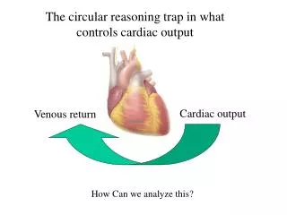
The volume of blood pumped by a ventricle per minute is the cardiac output of that ventricle! How is CO calculated? Memorize! How do we get the ventricle ready to push blood out? Most ventricular filling passively occurs as blood drains from vena cava and pulmonary veins through atria and into the apex (down hill) by simple gravity and draining…..water pours out of a glass onto the floor by the same effect. End Diastolic Volume(EDV): ml of blood in ventricle after the “kick” Pre-systole (130 ml) • Mostly from passive filling during Vent and Atrial diastole, plus a small bonus from atrial systole (about 100 ml + 30 ml from kick) Isovolumetric Contraction: no volume change (hopefully) • AV Valves just closed and Semilunars are not yet able to open! • Volume in ventricle does not change but pressure on this 130 ml goes up until pressure in ventricle is greater than in the artery in the blood on other side of the semilunar valve!
VentricularEjection/StrokeVolume(SV): blood leaving vent. (70ml) “Ejection Fraction (EF)” EF=SV/EDV X 100 = 70ml/130ml X 100 = 54% SV can be larger and EF can be up to 90% during exercise! End Systolic Volume (ESV): Amount left in vent. After systole ESV=EDV-SV=130ml-70ml =60 ml (amt. remaining during isovolumetric relaxation) Heart disease= a weak ventricle so the EF is small and ESV large Cardiac Output (CO): amount pumped by a ventricle/minute CO= SV X HR = 70 ml/beat X 75 beats/min = 5,000 ml/min CO=SV X HR = 100 ml/min X 50 beats/min = 5,000 ml/min (X2 for “whole heart”: Rt/Lt ventricles pump equal volumes: 5X2=10 L/min) Range (cardiac reserve): CO can get up to 35 L/min/Ventricle for Olympic Athletes! Your Blood volume (4-7 L) must be recycled many times/min at 35 L/min
Will I need to do the math for the calculation we just looked at on a lab or lecture test? Answer: YesWill I get to use a calculator: No-Will math be “easy”:Yes • Formulas should be understood for what they are….a logical prediction of what the body does. • Know basic formulas we just looked at. • Know normal values we just looked at. • Know how to calculate missing information given a set of known facts. • KNOW HOW AND WHY WE MODIFY C.O.!
Put ECG, Aorta, Ventricle, Atria, BP, heart sounds and pressure all together to measure cardiac output. If the RR-interval is 1.0 seconds, what is the aortic BP, SV and CO? VIP Diagram
SA Node-->(Atrial depolarization/contraction)AV NodeBundle of HisLt/Rt Bundle BranchPukinjeMyocardium (Depol./Contract)
ACTION POTENTIAL IN INDIVIDUAL CELLS vs. DEPOLARIZATION OF ENTIRE HEART (MANY CELLS CONNECTED BY GAP JUNCTIONS) . HUGE Functional Difference: Pacemaker Cell vs. Contractile Cell SA NodalCell(AV nodal or conduction)=(no contractile force) Cardiac Myocyte (force generation=contractile) Speed of Na+ leak determines rate of depolarization for SA/AV-Nodes Very SteepDepolarizes FastRapid Heart Rate Not Steep: Cells depolarize slowly take more time to reach threshold for voltage gated Na+ channels to open Slow Heart Rate Absolute refractory period determines rate of depolarization by determining time the cell must wait until it can be depolarized again Myocytes and Timing: Why a delay in the onset of contraction? • Na+ channels must open and let Na+ into cell • Ca++ gets inside myocytes • Ca++ must find troponin and pull it off actin • Actin must find/bind myosin • Actin must ratchet across myosin and generate force • Ca++ Slow to exit myocytes • These all add to the x-axis (time) of contraction • This generates force in ventricle: Isovolumetic Contraction Ejection
SA Nodal cells are non-contractile and rhythmically depolarize themselves and adjacent cells. Nodal cells eventually depolarize adjacent contractile cells via gap junctions. As a result, the depolarizations of contractile and non-contractile cells are very different! Non-Contractile Cells! Remember: this is depolarization in single cells, not the whole heart!
Agents like epinephrine speed up the heart rate and the parasympathetic NS (ACH) slows the heart down. What happens to the rate of membrane leakage (pacemaker potential) to accomplish this change in heart rate at the SA Node? Before and after Sympathetic Stimulation +pacemaker potential Longer period between depol Shorter period between depol Longer time to next depolarization Stimulation of the parasympathetic NS slows down the heart rate by hyperpolarizing the SA node and decreasing its leakiness (pacemaker potential)
Cardiac Output = Stroke Volume (ml/beat) X Heart Rate (beats/minute) Normal Person has a cardiac output of about 5 liters/min at REST SV=70ml/beat HR=70 beat/min SV X HR= 4,900 ml/beat (approx. 5 L/min) Cardiac Output in an aerobically fit Olympic Athlete?? Peak HR 170 beats/min Peak SV 160 ml/beat CO= 27.2 L/minute! So that means the 8 liters in a 155 lb person is recycled about 3-4 times/minute! In Heart Failure Patient (very large EDV, small Ejection Fraction)? HR= 80beat/min SV= EDV (190ml)-ESV (140 ml) SV=50ml/beat CO= 80 beats/min X 50 ml/beat = 4.0 L/min-perhaps even lower With respect to Hemoglobin oxygen extraction ration how are they still alive? Is their heart using more or less O2 to push more or less blood? See a problem? MESSAGE?? CO, HR, SV, and Oxygen Delivery are DYNAMIC!
FRANK STARLING LAW OF THE HEART: “Energy of contraction is proportional to the initial length of the cardiac muscle fiber”. This explains how a heart can regulate its own output based simply on venous return to the heart and conditions in heart. Normally the heart contracts at less than the ideal actin/myosin length and preload! Curve: EDV (ml) vs Stroke Volume (ml) However as the EDV is increased, the myocardium is stretched and the actin/myosin orientation becomes closer to ideal (In healthy heart!) Each “curve” is unique to conditions around heart: temp, CO2, etc. As EDV↑…Contactility↑…SV↑….therefore CO goes up…SO WHAT? • Heart moves more blood/beat for about the same ATP cost • Cardiac Output can increase with no change in rate • Adaptation can occur independently of nervous system • VIP: Limits to System Exist • With respect to cardiac reserve, why does your heart normally work/function in “middle” of ascending part of curve, and not at the top or back side of the curve?
The key is to ask where you are on the curve given the observed EDV for a given systole, this lets you predict the SV for that systole: Often Folks call this the ventricles “PRELOAD”
Frank-Starling curves are shifted by: A “Shift” makes the heart pump a larger or smaller stroke volume. A “Shift” creates a new F-S curve with a new shape. Factors That Modify the “Shape” of F-S curves: Make myocardium contract more/less forcefully • Sympathetic stimulation: • Digitalis and calcium uptake inhibitors: • Hypercapnia/Hypoxia- • Body temperature- • Thin myocardial walls (alcoholic heart)- • Many more factors exist…… • What determines where your heart is “Located” on a F-S Curve: • Answer: Preload and Stroke Volume
The original F-S Curve is modified based changes to myocardial function caused by temperature, positive ionotropic drugs, myocardial infarct,…etc.
Frank Starling Curve Shifts Up when the heart needs to work harder ( Add Epinephrine) and Shifts Down when the heart starts to fail (add Hypoxia or have an Infarct). If Preload Changes on a Single Curve Stroke volume changes.
WHAT HAPPENS TO THE HEART WHEN IT FAILS? In terms of the FS-curve, why will SV and cardiac output plummet? 1) Loss of myocardium due to infarct- Can you generate force if the myocytes are dead? 2) EDV gets too large- ejection fraction small 3) Actin/myosin are over stretched- no traction 4) Contractility is reduced and SV is reduced- This is bad! 5) Cardiac Output Plummets! Can you supply the heart with oxygen without CO? Other Classic Problems: • If heart hypertrophies (grows to big/thick)- • If heart dilates (creates a large EDV, small SV and a thin wall)- • During lung problems: emphysema, core pulmonare, etc…. • Problem:Law of Laplace: ^radius…^tension…^^^^work • These examples can result in death from a loss in cardiac reserve and a set or neurological responses that make the original problem WORSE and lead to a heart attack.
A failing heart results in the accumulation of fluids (edema) in the lung or body. With respect to Rt or Lt ventricular failure, where does the pressurized fluid accumulate? Why? What could cause Rt/Lt Failure? Why might your brain try to make the heart work even harder? Can it work harder?
WHAT ARE SOME MECHANISMS FOR IMPROVING VENOUS RETURN IN A LOW PRESSURE (VENOUS) LOOP? • Simple pressure gradient-Systemic vs. Pulmonary circuits mmHg mmHg • Thoracic (Respiratory) Pumping by ribs/sternum • Valsalva Maneuver and preload changes: heart attack risk • Cardiac Suction: Negative Pressure in Atria or Ventricles • Venous Return to Heart: Venous valves and skeletal muscle activity- • Varicosities and Anal Hemorrhoids • Gravity changes pressure: ±1.92 mmHg/inch of elevation or depression • If the top of your head is 15 inches higher than your head the BP at the higher location would be decreased by 29 mmHg(15X1.92= -28.8mmHg) • BP at brain is: (120-29)/(80-29)=91/51 • BP at foot is added! 40X1.92=77 mmHg 199/157 • If you are dizzy, why does laying down prevent you from passing out? • Why does your sprained ankle hurt and swell more if you are standing up?
Gravitational effects on blood flow and pooling are part of the reason we are asked to lay flat when we feel dizzy or why our sprained ankles “throb” and become swollen only when we are standing up.Remember:+/- 1.92 mmHg pressure for each Inch of elevation or depression!If the brain has no pressure, there is no blood being delivered!
Venous pumping provides a very simple and very effective way to increase the return of venous blood to the heart? (If venous return to atria causes the Preload to increase too!)
How do we integrate changes in pressure, volume, ecg, EDV, ESV, SV, CO, heart sounds and time, with the 5-step cardiac cycle?Why is this clinically important to you?How would changes in the function of the nodes, BB, and valves make this look different?