Download
1 / 58
580 likes | 746 Views
Immunology 146:474. Tu, Fri 1st period (8:40-10:00 AM) Serc 118 Dr. Lori Covey-Office hrs: 1-3 Thursday Dept. of Cell Biology & Neuroscience Nelson Hall, B314 covey@biology.rutgers.edu Class web site: http://lifesci.rutgers.edu/~covey/Immuno/index.htm. MHC Class I structure.

E N D
Immunology 146:474 • Tu, Fri 1st period (8:40-10:00 AM) • Serc 118 • Dr. Lori Covey-Office hrs: 1-3 Thursday • Dept. of Cell Biology & Neuroscience Nelson Hall, B314 • covey@biology.rutgers.edu • Class web site: http://lifesci.rutgers.edu/~covey/Immuno/index.htm
MHC Class I structure b-pleated sheet b-pleated sheet
Figure 3-23 NH2 terminus NH2 terminus COOH terminus
Peptides eluted from two different MHC class I molecules Green residues are the anchor residues, Y=tyrosine, F=phenylalanine, both aromatic amino acids
Peptides that bind MHC class II molecules are variable in length Peptides are at least 13 amino acids long and can be longer
Specificity of a T cell receptor is defined both by the peptide it recognizes and by the MHC molecule bound to it
Figure 1-24 part 2 of 3 Fc receptors
Figure 1-15 Macfarlane Burnet theory of clonal selection-1950s
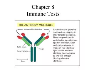
Mature B cells display membrane-bound Immunoglobulin (antibody) molecules, which serve as receptors for antigen. Each molecule of Ab on the membrane of a single B cell has an identical binding site for antigen. Bone marrow, pre-B cells
Interaction between antigen and the membrane-bound antibody (mIg), as well as interactions with T cells and macrophages, selectively induces the activation and differentiation of B-cell clones of corresponding specificity. Selected B cells divide repeatedly and differentiate over a 4-5 day period generating a pool of plasma cells (terminally differentiated) and memory cells. 2o lymphoid organs Germinal centers
To activate a B cell, it needs to receive a signal from antigen recognition through the mIg and a signal from CD4+ T cells
Mature B cells display membrane-bound Immunoglobulin (antibody) molecules, which serve as receptors for antigen. Each molecule of Ab on the membrane of a single B cell has an identical binding site for antigen. Bone marrow, pre-B cells
Mechanisms for generating diversity Number of total number of antibody specificities available to an individual is known as the antibody repertoire Ig genes are rearranged in antibody-producing cells Process called “Somatic recombination”
A single B cell gives rise to a single antibody molecule with two identical heavy and two identical light chains A resting, naïve B cell expresses IgM and IgD on its surface as its BCR (B cell receptor).
Southern blot analysis of V region and C region. Digest DNA with restriction enzyme and separate pieces of DNA in an electrical field. Fragment of DNA Labeled RNA representing Bence-Jones Protein (light chains) Hybridized to DNA isolated From either fibroblasts or B cells
Organization of the Human Heavy chain variable region loci VH region The Variable region of the Ig heavy chain is encoded by three Gene segments; The V segment 95-101 amino acids The D segment 2-5 amino acids The J segment up to 13 amino acids
Organization of the Light Chain Loci The Variable region of the Ig light chain is encoded by two Gene segments; The V segment 95-101 amino acids The J segment up to 13 amino acids
Figure 4-3 The heavy and light chain genes are located on autosomes so there are 2 copies of the heavy chain locus, 2 copies of the light chain and light chain loci Possibility of generating 1011 antigen specificities
First mechanism that generates diversity Combinatorial Diversity Any functional VH segment can recombine with any DH segment and any JH segment Any functional VL segment can recombine with any JL segment
D-->J rearrangements on both alleles VH DH JH C V-->DJ rearrangement VHDJH C
Figure 3-6 Framework and hypervariable regions define the Variable Regions of the Ig heavy and light chain polypeptides
12/23 7-mer 9-mer Recombination Recognition Sequences (RSS) Only RSS elements that have different spacer lengths can recombine with each other
12 or 23 7-mer 9-mer Recombination Recognition Sequences (RSS) Only RSS elements that have different spacer lengths can recombine with each other
Figure 4-7 Proteins required for carrying out recombination called “V(D)J recombinase RAG I and II enzymes required For variable region Somatic recombination Create hairpins Which are cleaved. Terminal deoxynucleotidyl transferase (TdT) are added, nt can be subtracted as well by nucleases
Figure 4-7 part 3 of 3 2nd mechanism for creating diversity: Junctional Diversity Enzymatic processes creates Diversity in the joint between gene segments
Figure 4-8 part 1 of 3 Creates tremendous diversity at the joins of the three segments
Figure 4-7 part 3 of 3 3rd mechanism for creating diversity: Combinatorial- at the heavy and light chain level Pairing of individual heavy chains with different light chains
Somatic hypermutations create diversity • Occurs later after B cells are in secondary lymphoid organs • Introduces single nt mutations into germline DNA • Results in increased binding affinity of antibody for antigen
4th mechanism for creating diversity: Somatic hypermutation Occurs after stimulation with antigen in the antigen-dependent stage of B cell differentiation Introduces point mutations into the V regions of the rearranged heavy- and light-chain genes at a very high rate Requires a signal from CD4+ T cells