Download
1 / 47
540 likes | 1.36k Views
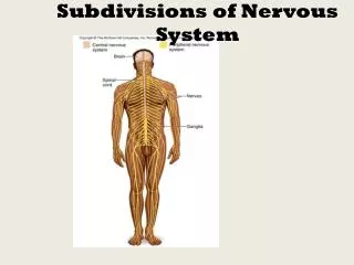
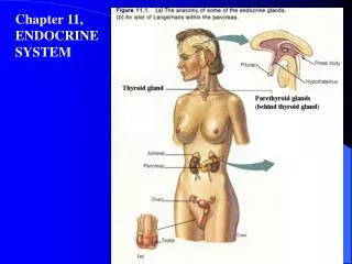
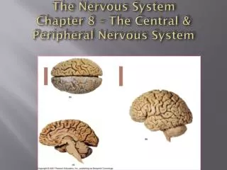
The Nervous System Chapter 8 – The Central & Peripheral Nervous System. Central Nervous System (CNS). Brain enclosed within cranium -Comprised of four major regions: Cerebrum, Cerebellum, Diencephalon, Brainstem

E N D
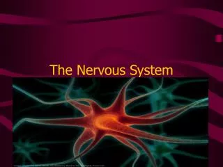
The Nervous SystemChapter 8 – The Central & Peripheral Nervous System
Central Nervous System (CNS) • Brain enclosed within cranium -Comprised of four major regions: Cerebrum, Cerebellum, Diencephalon, Brainstem • Spinal Cord begins at foramen magnum, runs through vertebral foramen (spinal canal), and ends at L2 vertebral level by forming conusmedularis • CNS is well protected by bones, CT meninges, and fluid (cerebrospinal fluid (CSF))
Meninges Membranes that surround and protect the CNS • Three layers: • Dura mater • Arachnoid mater • Pia mater
Dura Mater – tough, fibrous outer layer; • 2 layers thick around brain with creation of dural sinuses between layers; • 1 layer around spinal cord with epidural space external
Arachnoid– “spidery” web-like middle layer • Pia Mater – delicate, thin inner layer; extension of pia mater (“filumterminale”) extends from tip of cord to coccyx to anchor cord in place Subarachnoid space – between arachnoid & pia mater; contains cerebral spinal fluid (CSF) • sample of CSF can be taken at subarachnoid space inferior to the conusmedularis by “lumbar puncture” (spinal tap)
The Spinal Cord Begins at foramen magnum & ends at L2 vertebral level by forming conusmedularis • Has 2 thickened areas • cervical enlargement - supplies nerves to upper extremity • lumbar enlargement - supplies nerves to lower extremity (conus medularis) Made up of 31 spinal cord segments
Dorsal root ganglion (DRG) Dorsal root Ventral root • Each spinal cord segment has a pair of • dorsal roots with their associated dorsal root ganglia (DRG) • ventral roots
Each dorsal root contains the axons of sensory neurons (unipolar neurons) • Each dorsal root ganglion contains the cell bodies of these sensory neurons • Each ventral root contains the axons of motor neurons (multipolar neurons whose cell bodies are within the cord)
The dorsal & ventral roots of each segment come together at the intervertebral foramen (IVF) to form a mixed spinal nerve
Spinal Nerves • Part of the PNS • Contain both motor & sensory fibers (“mixed nerve”) • 31 pair of nerves – each nerve forms from union of dorsal/ventral root of spinal cord segment & exits between vertebra at IVF (intervertebral foramen) • 8 pair cervical spinal nerves – 1st cervical nerve exits between occipital bone & C1, 8th cervical nerve exits the IVF between C7-T1 • 12 pair thoracic spinal nerves • 5 pair lumbar nerves • 5 pair sacral nerves • 1 pair coccygeal nerves
Below the conusmedularis, spinal nerves must angle downward (in the subarachnoid space) before exiting their IVF. These spinal nerves make up the caudaequina Cauda equina
Spinal Nerves • Once formed, spinal nerves will branch • The branches of most spinal nerves (comprised of axons) interweave to form nerve plexuses • peripheral nerves then branch from the plexuses to provide motor & sensory innervation to specific areas of the body
Nerve Plexus • 4 major plexuses • cervical • brachial • lumbar • sacral
Spinal Nerve plexuses • Cervical plexus (C1-C5) • gives off phrenic nerve • Brachial plexus (C5-T1) • gives off median, ulnar • & radial nerve • Lumbar plexus (T12-L4) • gives off femoral nerve • Sacral plexus (L4-S4) • gives off sciatic nerve • No plexus forms between T2-T11 – intercostal nerves
Posterior median sulcus Posterior column Posterior gray horn - sensory Central canal Lateral column Lateral gray horn (T1-L2, S2-S4) - autonomic Anterior gray horn - motor Anterior column Anterior median fissure Sectional Anatomy of the Spinal cord Gray commissure
The spinal cord has a narrow central canal surrounded by “horns” of gray matter connected by a commissure. Gray matter horns contain sensory & motor nuclei (groups of cell bodies). Gray matter is surrounded by white matter “columns” which are made up of groups of myelinated axons creating organized ascending & descending tracts.
Tracts(Sensory & Motor Pathways) • Groups of axons found in the white matter columns of the spinal cord that carry specific information • Ascending tracts - carry sensory information up the spinal cord to areas of the brain • Descending tracts – carry motor information from the brain down to specific levels of the spinal cord • Ascending & descending tracts within the spinal cord are part of the sensory & motor pathways of the nervous system
Ascending Tracts (sensory pathways) • Spinothalamic tracts • carries poorly localized touch, pressure, pain & temperature from cutaneous receptors to the thalamus • from thalamus, some of this sensory info reaches primary sensory cortex of the cerebrum for “interpretation” & conscious awareness
Ascending Tracts (sensory pathways) • Posterior Columns • carries highly localized discriminative (fine) touch, vibration, conscious proprioception (position sense) to nucleus in medulla oblongata (M.O.) • from M.O., info travels along rest of pathway to thalamus & then to primary sensory cortex of cerebrum
Ascending Tracts (sensory pathways) • Spinocerebellar • carries proprioceptive (positional) information to the cerebellum (unconscious awareness)
Descending Tracts (motor pathways) • Corticospinal (pyramidal) • carries commands from primary motor cortex of cerebrum for conscious (voluntary) control of skeletal muscles. • most fibers cross in “pyramidal decussation” of medulla oblongata so that left cerebral cortex controls muscles on right side of body, & vice-versa.
Descending Tracts (motor pathways) • Medial & lateral pathways • originate from a variety of brain nuclei & send signals to motor neurons in the spinal cord for (subconscious) coordination of skeletal muscle activity, maintenance of posture & muscle tone.
Ascending & descending tracts are part of larger sensory & motor pathways • These sensory & motor pathways include the afferent & efferent neurons of the PNS • Sensory & motor information gets in/out of spinal cord via spinal nerves
The Brain • Brain stem • medulla oblongata (M.O.) • pons • midbrain • Diencephalon • thalamus • hypothalamus • epithalamus (pineal gland) • mamillary body • Cerebrum • Cerebellum Cerebrum T P P H midbrain M Cerebellum pons m.o.
Cerebrospinal Fluid (CSF) • clear, colorless fluid formed by filtration of blood plasma by choroid plexuses within ventricles of the brain. • circulates through ventricles, into central canal of spinal cord & around brain & SC in subarachnoid space. Reabsorbed through arachnoid granulations into dural sinuses & then into bloodstream • functions in protection of CNS, support, nutrient supply, waste removal • sample of CSF can be taken at subarachnoid space inferior to the conusmedularis by “lumbar puncture” (spinal tap)
The Brainstem • Medulla oblongata • continuation of the SC above the foramen magnum • contains the pyramidal decussation • cranial nerve nuclei (XII-VIII (cochlear) • cardiac, vasomotor, & respiratory reflex centers • Pons • “bridge” linking cerebellum to SC & other parts of brain • cranial nerve nuclei (VIII (vestibular) – V) • respiratory center
The Brainstem • Midbrain • cerebral peduncles – location of descending (motor) tracts • corpora quadrigemina • superior colliculi – visual reflex centers • inferior colliculi – auditory reflex centers • cranial nerve nuclei (IV-III) • reticular formation – network of interconnected nuclei throughout brainstem responsible for maintaining states of consciousness
The Diencephalon • Thalamus • surrounds 3rd ventricle • 2 halves connected by intermediate mass • comprised of sensory nuclei • functions primarily as a sensory relay station
The Diencephalon Hypothalamus • connects to pituitary gland via the infundibulum • has many important functions relating to maintaining homeostasis including: • integrating nervous & endocrine systems through control over pituitary gland • integration of ANS from visceral stimuli • hunger/satiety, thirst, body temp. regulation • hormone production (ADH, oxytocin) • subconscious coordination of motor responses associated with rage, pleasure, pain, sexual arousal • Mamillary bodies– reflex centers associated with eating, & processing of olfactory sensations
The Diencephalon • Pineal gland • secretes Melatonin which helps regulate day-night cycles (circadian rhythm)
Limbic system • functionally related areas in cerebrum, thalamus & hypothalamus involved in • emotional states & behaviors • linking conscious areas of cerebrum with unconscious areas of brainstem • long term memory
Cerebrum gyrus sulcus
Lateral sulcus (Insula is deep to lateral sulcus) Lobes of Cerebral Hemispheres Central sulcus Parietal lobe Parieto-occipital sulcus Frontal lobe Occipital lobe Temporal lobe
Gray & White matter of cerebrum • Gray matter – outer cortex & inner nuclei (centers) • White matter – deep to cortex; comprised of fibers (pathways for communication): • association • commissural • projection
White matter of cerebrum Association fibers • association fibers – connect gyri in same hemisphere • commissural fibers – connect gyri in opposite hemispheres (e.g. corpus callosum) • projection fibers – connect cerebrum with other parts of brain & spinal cord Commissural fibers Projection fibers
Functional areas of Cerebrum • Motor and Sensory areas - receive sensory info & generate motor (skeletal muscle) responses • Association areas – interpretation of sensory info & planning and coordination of motor responses • Cerebral processing centers - higher order integrative & analytical functions
Motor & Sensory primary motor cortex (precentral gyrus)
Motor & Sensory primary sensory cortex (postcentral gyrus)
Motor & Sensory primary motor cortex (precentral gyrus) primary sensory cortex (postcentral gyrus) gustatory cortex visual cortex auditory cortex olfactory cortex
Association areas • interpret incoming sensations; coordinate motor responses somatic motor association area (premotor cortex) visual association area
Cerebral Processing Centers • higher-order integrative centers • may be unilateral general interpretive area (Wernike’s) –Lt hemisphere usually motor speech center (Broca’s) - Lt hemisphere usually Prefrontal cortex (bilat.)
transverse fissure arbor vitae (white matter) folia (gray matter) The Cerebellum • 2 hemispheres connected by vermis • separated from cerebrum by transverse fissure • outer folia with inner arbor vitae • functions include control of skeletal muscles (unconscious) for balance, coordination & posture • Stores patterns of movement • links to brainstem by cerebellar peduncles
Cranial Nerves • 12 pairs of nerves (part of PNS) that connect to the brain; provide motor, sensory &/or autonomic (parasympathetic) function
Cranial Nerves (know #, name & basic function) I Olfactory – smell II Optic – sight III Oculomotor– motor to eye muscles; ANS for accommodation of lens & pupil constriction IVTrochlear – motor to one eye muscle VTrigeminal – motor to muscles of mastication, sensation to face & mouth VIAbducens– motor to one eye muscle VIIFacial – motor to muscles of facial expression; taste; ANS to lacrimal & salivary glands VIIIVestibulocochlear– equilibrium & hearing IXGlossopharyngeal – swallowing, taste, ANS to salivary glands, sensory reception from monitoring of blood pressure in large arteries XVagus– sensation from viscera; ANS visceral muscle movement (respiratory, digestive, cardiovascular systems) XIAccessory – motor to muscle of pharynx, SCM & Trapezius XIIHypoglossal – motor to tongue muscles