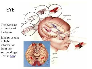
EYE PROJECT
EYE PROJECT. Austin Petty Travis Byrne Period 6. Table Of Contents. Parts of Eye & Function Dissection Lab Disorders of Vision & Treatment Diseases of Eye & Treatment Neural Pathways of Vision & Perception in Brain Study Questions Interview With Doctor Zuzana R. Gellner

EYE PROJECT
E N D
Presentation Transcript
EYE PROJECT Austin Petty Travis Byrne Period 6
Table Of Contents • Parts of Eye & Function • Dissection Lab • Disorders of Vision & Treatment • Diseases of Eye & Treatment • Neural Pathways of Vision & Perception in Brain • Study Questions • Interview With Doctor Zuzana R. Gellner • Field Of Vision • Visual Illusions • The Cookie Page!! • Bibliography
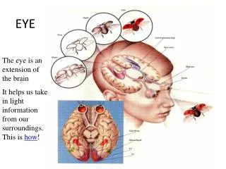
Parts & Functions of Eye three major layers of eye: outer- fibrous tunic cornea major role in focusing bends (refracts) light sclera protection & attachment of eye upholds the shape of eye middle- vascular tunic iris regulates amount of light to retina ciliary body releases aqueous humor transforms frame of lens choroid takes in light blood vessels provide eye tunics
Parts/Function Continued inner- retinal tunic pigmented layer accumulates vitamin A sucks in light nervous layer catches light converts light into- action Six extrinsic muscles of eye:
Parts/Functions Continued Retina: pigmented layer accumulates vitamin A sucks in light nervous layer catches light converts light into action photoreceptors contains rods and cones rods more than 100 million in each eye enhanced by low light enhanced by shapes and movement can’t separate small details cones- cone shaped cells distinguish fine details about 7 million in each eye high density in posterior center of retina less sensitive fovea centralis most precise vision no rods macula lutea yellowish lots of cones
Parts/Functions Continued optic disk where optic nerve (CN II) exits eye no photoreceptors ---> BLIND SPOT optic nerve (CN II) forms with axons of ganglion neurons Conjunctiva- mucus membrane over inner face of eye lids lubricated moist palpebral conjunctiva thick inner surface bulbar (ocular) conjunctiva thin anterior surface superior & inferior conjunctiva sacs both defend eye from materials entering eye medication goes here often PINKEYE-conjunctivitis bacterial virus dry & scaly in need of vitamin A highly contagious
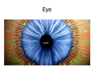
Parts/Functions Continued Cornea major role in focusing bends (refracts) light abundant nerve supply stimuli causes blinking increase secretion-lachrymal glands tough - - 5-layered membrane Iris between cornea & lens circular shape inside circular muscle radial muscle color in eyes due to amount of pigment high-appear brown or black low-appear blue,green, or gray Pupil center hole in iris light is directed through circular muscles gets small in bright light/seeing close things gets big in dim light/seeing far away things
Parts/Functions Continued Lens biconvex structure-flattened sphere held in by ligaments easily bendable change in shape due to ciliary muscles focusing &distance adjustments transparent fibers Ciliary Body ciliary muscles change shape of lens (focusing) ciliary process makes aqueous humor (clear watery fluid) Suspensory Ligaments attaches lens & ciliary body Anterior Cavity two chambers- anterior chamber between iris & cornea full of aqueous humor posterior chamber between suspensory ligaments & iris full of aqueous humor
Parts/Functions Continued Choroid Plexuses secretes aqueous humor into posterior chamber Aqueous Humor provides oxygen & nutrients to lens & cornea Canal of Schlemm pipes aqueous humor from anterior chamber into blood stream Vitreous Humor - jelly like fluid fills posterior cavity passes light to retina keeps up intraocular pressure Protective Structures: eyebrows just above eyes on outside keep moisture from running into eyes perspiration (sweat) oily fluids shading eyes prevents materials from superior contact
Parts/Functions Continued eyelids (palpebrae) close over eyeball act as screen to keep particles out of eye eyelashes protect eyes from particles in air nerve endings touched ---> cause closing of eyelids tear glands lubricates eye when closed washes matter from eye’s surface *FACT* - Humans blink about every 6 seconds
Dissection Lab Step 1- Have your dissection tools and tray out in front of you Step 2- Remove your eyeball from the air-tight plastic carefully Step 3- Next, take your eye ball and examine it and try to get an understanding of what you are dissecting *remember you won’t know if your eye ball is the left or right Unless all ready specified. Step 4- Now, remove all of the fatty tissues (the white and yellowish stuff) carefully not to cut any muscles or other valuable things. Remember, you are trying to keep all of the parts together and connected the eyeball, so don’t cut anything off except for the fatty tissues. Keep the cut fatty tissues in your dissection tray for now. * a trick in removing the fat is to make small insertions and to look for creases in the eye ball where the fatty tissues meet with other parts of the eye. Such as muscles. Step 5- After you have taken most, if not all of the fat off, you should have your six extrinsic muscles. Now you will be able to figure out if your eye ball is a left or right by the oblique muscles (superior and inferior for they go towards the medial aspect of the body. Step 6- Now find the back of the eyeball and you will find and area where it comes out.here you will cut into the eye ball to find all of the vital parts inside of the eye. Take your razor and cut carefully and slowly downward make a nice clean insertion. You will come to a hard texture which will be the sclera. Here you will stop and try to separate the eye carefully keeping it all intact. Step 7- Once you are inside of the eye, you will find all of the internal parts of the eye. Step 8-You can come and ask us any questions you have concerning this dissection.Anything at all.Now go have fun!
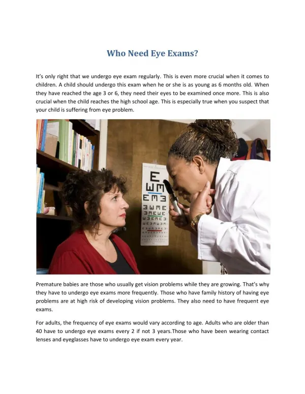
Disorders Of Eye Myopia- nearsightedness Description: elongated shape of eye, improper refraction of light (focus to early) Cause: inherited --detected between ages 8-12 Treatment: glasses, contacts, & REFRACTIVE SURGERY- serious cases Hyperopia- Farsightedness (first form) Description: ease in seeing distance & hard seeing nearer objects Cause: eyeball is too short (light rays focus behind retina) Treatment: glasses Presbyopia- farsightedness (2nd form) Description:lens of eye loses flexibility Cause: aging Treatment: reading glasses Symptoms: vision diminishing, cant see object up close
Disorders of Eye Continued Watery Eyes Description: excessively watery eyes, lack of necessary substances to keep eye moisturized Cause: allergies, infections, small object lodged in eye Treatment: see ophthalmologist Dry Eyes Description: very common, more common in women, Cause: lack of tear production, allergic reaction to medications, tears can’t keep eye moist or at right p-h balance Treatment: replacing or conserving tears, lubricating ointments, SERIOUS CASES---> plug puncta to with- hold-tears Symptoms: burning of eyes, itching, excess tearing (sometimes) Crossed Eyes (in children)-- strabismus---> eyes inward (esotropia) eyes outward (exotropia) Description: misalignment, Cause: born with it, develop early in life, Treatment: see doctor when it comes up, earlier treatment, better the chance of treating Cataract Description: common, clouding of crystalline lens Cause: aging, injury, diabetes Treatment: surgery frequently performed around the world
Disorders of Eye Continued Drooping Eyelids Description: loss of muscle elasticity Cause: born with it, aging, allergic reaction, and other medications Treatment: surgery Symptoms: eyelid sags, if it impairs vision consult a health care provider for further information Astigmatism Description: irregular curvature of cornea (usually shaped like football) Cause: look above Treatment: contacts, glasses Symptoms: blurry vision, headache, eye strain Partial Color Blindness: Dichromatism Description: mostly in males, inability to distinguish colors (hard w/ red and green), most common Cause: defect in retina (other nerve portions of eye), heredity Treatment: none *TEST* - - - Given to public employees (police/firemen) normal will see 57 partial color blindness will see 35
Diseases of Eye Pink Eye - conjunctivitis Description: inflammation of conjunctiva Cause: bacterial or viral infection, allergy, irritated eye Treatment: anti-biotic or ointment prescription Symptoms: swelling, redness, irritation, yellow sticky discharge which causes eyelids to stick together Flaking Eyelids (blepharitis) Description: inflammation of eyelids Cause: oily skin, dandruff (of eyelids), or dry eyes Treatment: Medications & daily cleansing Symptoms: irritation, itching, rarely red eye Histoplasmosis this disease is thought to have been spread from the lungs to the eye by a bacterial virus known as Histoplasma Capsulatum spores which cause a mild inflammation Blepharitis (swollen eyelids) Description: inflammation of eye lids & lashes Cause: excessive oil on eyelid, bacterial infection, allergic reaction, poor eyelid care Treatment: see Optometrist
Diseases of Eye Continued Chalazion Description: inflammation of meibomian gland (in eyelid) Cause: blockage of duct to eyelid surface---->it may be an infection Treatment: soak in warm water, 6+ weeks surgical removal Symptoms: pain, tenderness of eyelid, conjunctivitis Dacryocystitis Description: inflammation of lachrymal sac Cause: not really known Treatment: rub with warm pad, ointments, medication Symptoms: swelling & redness at nasal side of eye Ocular Albinism Description: Lack melanin pigment Cause: Inherited Treatment: visual aids, Change in living conditions, surgery (not real effective) Retinoblastoma Description: rare, Causes malignant tumors --> retinal layer of eye Cause: hereditary Treatment: Removal of eye- if untreated could be fatal
Neural Pathway & Perception Neural Pathway : takes sensory information to brain Axons the long, tubular extension of a neuron that transmits nerve impulses away from the cell body Horizontal Cells transmits info laterally nervous layer of retina help in color seeing increases input of moving info to brain Amacrine Cells increase changes in illumination of retina Optic Chiasma axons cross over axons enter optic tracts Optic Tracts axons from lateral (ipsilateral) & medial (contralateral) has information from both halves of visual fields
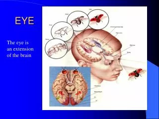
Neural Pathway & Perception Continued Lateral Geniculate Nuclei thalamus Visual areas of the Occipital Lobe cerebrum information ends up here on visual fields Steps: photoreceptor---> bipolar neuron---> ganglion neuron---> optic nerve (CNII)---> optic chiasma---> optic tract---> lateral geniculate---> visual cortex of occipital lobe Perception: Optic Nerves give messages from retinas to lateral geniculate nuclei of thalamus *Messages must be sent to both the primary visual areas and visual association areas for images to be scene *complex things are interpreted in the temporal association areas *when the primary visual areas are messed up, it results in blindness (the failure of seeing) *in posterior portion of temporal lobe printed words may be made out
Study Questions • Parts and Functions 1. Name the six eccentric muscles of the eye 2. The retina has two layers. Give those layers and their functions. 3. What is the difference between cones & rods? • Disorders of Vision 1. What is the difference between nearsightedness and farsightedness? Why? 2. What causes Astigmatism? 3. Some of the symptoms of this disorder are itching, burning of the eyes and sometimes excess tearing. • Diseases of eye 1. Name two diseases the inflames the eye and tell some information about those diseases. 2. ________________ is a disease that inflames the lachrymal sac. 3. This is an inflammation of the conjunctiva.
Study Questions Continued • Neural Pathways 1. Write down the path of the axons. 2. What are two types of cells that help transmit information in the retina? 3. When the information gets to the visual areas of the occipital lobes, what information is there? • Dissection 1. Can you identify if your eye is either a left on or a right one? What do you need to do first to make sure? 2. Did you make big insertions or small insertions? Why ? 3. What is the color of fat? Muscle? 4. When you make your insertion into the eye, what is hard texture that you come upon? What color is it? 5. What is the part that is greenish-bluish? What is the function for that? 6. What is the jelly-like substance inside of the eye?
Interview With Doctor Gellner • See Video of Interview and Answer Following Questions: Questions- 1. Are glasses more common than contacts? Why? 2. Name one technological advance in contacts. 3. According to Dr. Gellner, what is the most common eye disorder she comes across? 4. What is the first thing that Dr. Gellner does before a patient sees her for an appointment? 5. What instrument measures a person’s vision? 6. What instrument does Dr. Gellner use to measure the shape of an individual’s eye? What is the measuring for? 7. What is the name of the microscope that Dr. Gellner uses?
Field Of Vision This is a diagram of your field of vision. The blue and dark purple represent where you can see with your left eye. The light purple and dark purple represent what you can see with the right eye. Dark purple shows your field of binocular vision. Why do you think that with binocular vision you see a smaller area?
Visual Illusions (1) Are the two lines intersecting? What do you think? (2) Which like segment is longer? What do you think? Answers: (1) the line segments do not intersect. (2) The line segments are the same
Visual Illusions Continued (3) This is a lithograph that was drawn by M. C. Escher and represents Einstein’s theory of relativity “perception changes depending upon one’s position and motion.”
The Cookie Page!!! *Refer to the cookies Statistics - Six out of every ten people have Brown eyes Two out of every ten people have Green eyes Two out of every ten people have Blue eyes *This shows that the majority of people in the world have brown eyes. This goes to show that the color brown in eyes is the DOMINATE GENE.* Enjoy!!!!
Bibliography • Human Anatomy & Physiology - Second Edition - Solomon - Schmidt - Adragna - copyright 1990 by Saunders College Publishing • Encarta 1995 - CD • www.encarta.msn.com • Grolier's Encyclopedia - CD • Your Human Body - CD • Canadian Ophthalmological Society • www.I-care.net • Eye Clinic Surgery Information • www.rochestereyecare.com • yahoo.com • www.intelihealth.com • www.nei.nih.gov/ • Glaucoma Research Foundation - Rita Loskill • John Hopkins Health Information • The Question & Answer CD About the Human Body • http://www.eyesite.com/Eye_Problems/eye_problems_disease.htm