Download
1 / 1
10 likes | 174 Views
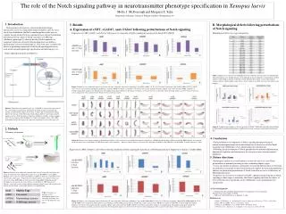
The role of the Notch signaling p athway in neurotransmitter p henotype s pecification in Xenopus laevis. Molly J. McDonough and Margaret S. Saha. Department of Biology, College of William and Mary, Williamsburg, VA. 3. Results.

E N D
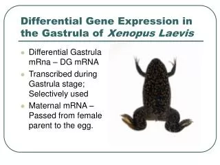
The role of the Notch signaling pathway in neurotransmitter phenotype specification in Xenopuslaevis Molly J. McDonough and Margaret S. Saha Department of Biology, College of William and Mary, Williamsburg, VA 3. Results B. Morphological defects following perturbations of Notch signaling 1. Introduction Injected Side A. Expression of xNBT, xGAD67, and xVGlut1 following perturbations of Notch signaling During primary neurogenesis, neurotransmitter phenotype specification occurs in a nonrandom dispersed pattern, and, due to its role in lateral inhibition, the Notch signaling pathway may serve to regulate this patterning. Previous experiments have shown that blocking downstream effecter genes of Notch can induce a GABAergic (inhibitory) phenotype (1) while by-passing Notch completely by exogenous expression of neural determination genes can induce a glutamatergic (excitatory) phenotype (2). This study aims to explore the effects of perturbing components of the Notch signaling pathway on early neurotransmitter phenotype specification and patterning in vivo. Expression of xNBT, xGAD67, and xVGlut1 following over expression of Notch signaling by injection of X-Notch ICD (xNICD) Morphological defects by stage and injection Internal control Notch signaling in primary neurogenesis A B C xNICDlow-dose (1.5ng) injection Table 2. Embryos were analyzed for morphological defects following fixation and in situ hybridization. Abnormal embryos were divided into one of two mutually exclusive categories. “Gross Defects” denotes embryos which failed to develop or that had severely deformed body plans. “Neural Tube Defects” includes embryos with misshapen neural tubes as well as embryos in which the neural tube failed to close properly. D E F Figure 3. Embryos were unilaterally injected at the two-cell stage with either 4.6nl water containing 1.5ng xNICD mRNA (A, B, C) or 9.2nl water containing 3.0ng xNICD mRNA (D,E,F). xNICD serves to over express Notch signaling by introducing exogenous NICD (see Figure 1). Embryos were fixed at late neurula through swimming tadpole stages and assayed for xNBT, xGAD67, or xVGlut1 using whole mount in situ hybridization. The graphs in this figure indicate the percentage of embryos in which a decrease in expression for the specified marker was observed on the injected side in comparison to the un-injected side which served as an internal control. xNICD high-dose (3.0ng) injection Whole mount analysis of expression of xNBT, xGAD67, and xVGlut1 using in situ hybridization Figure 1. Expression of a proneural gene (e.g. X-NGNR-1) activates the expression of the transmembraneligand Delta. Delta binds the Notch receptor on adjacent cells causing a conformational change in the Notch receptor. This conformational change induces the cleavage of the Notch intracellular domain (ICD) by presenilin-catalyzed gamma-secretase. ICD then binds to Supressor of Hairless (xSu(H)), and this complex drives the transcription of Hairy/Enhancer of Split transcription factors. These HES transcription factors then repress the expression of proneural genes in the target cell. 2. Methods Figure 6. Graphical representation of the data presented in Table 2. Each chart represents the percentage of Gross Defects and Neural Tube Defects (NTDs) observed for each stage studied with each injection dose. Schematic of methods Unilateral Microinjection 4. Conclusions Sense, capped mRNAs Figure 4. Embryos were staged according to Nieuwkoop and Faber (1994) prior to in situ hybridization and were cleared in 2:1 BB:BA for photography. xNICD and xSu(H) DBM embryos were injected on the left lateral side as determined by GFP fluorescence prior to fixation. * indicates embryos that were injected with a high dose of mRNA (3.0ng xNICD or xSu(H) DBM). Scalebar represents 1.5mm. • Early perturbation of components of Notch signaling disrupt patterning of neurotransmitter phenotypes but do not demonstrate an instructive role for Notch signaling in the GABAergic versus glutamatergic fate specification. • Following initial interruptions of Notch perturbations on neuronal differentiation, homeostatic regulatory mechanisms may be acting to restore a normal neuronal population. Normal Development Expression of xNBT, xGAD67, and xVGlut1 following inhibition of Notch signaling by injection of a DNA binding mutant of Suppressor of Hairless (xSu(H) DBM) Fixation 5. Future directions Whole mount in situ hybridization xSu(H) DBM low-dose (1.5ng) injection • Histological analysis of assayed embryos to study the effects of early Notch perturbations on neuronal patterning in later (swimming tadpole) stages • Assays for markers of apoptosis and markers of neuronal differentiation to determine the molecular mechanisms acting to homeostatically regulate the neuronal population in embryos in which initial perturbations of Notch resulted in an excess or deficiency of differentiated neurons • Exposures of cell cultures or explants to DAPT, a pharmacological blocker of Notch signaling, at older stages to transiently alter Notch signaling and study the effects of these alterations more specifically on the GABAergic versus glutamatergic fate specification. Figure 2. Embryos were unilaterally injected at the two-cell stage with 4.6nl water with 1.5ng or 9.2nl water with 3.0ng X-Notch ICD (3) or xSu(H) DBM (4) sense mRNA. Embryos were co-injected with 0.5 ng GFP sense mRNA to allow for visualization of the injected side at later stages. Vehicle-injected control embryos were injected with 4.6 nl water with 1.0 ng GFP mRNA or 9.2nl water with 2.0ng mRNA. Following normal development, the injected side of the embryo was identified by GFP fluorescence. Embryos were fixed at early neurula through swimming tadpole stages and assayed for the markers listed in Table 1 using whole mount in situ hybridization. Labeled, antisense mRNA probes A B C Table 1.Summary of antisense in situ hybridization probes used indicating gene name and type of cell marked by each probe. xSu(H) DBM high-dose (1.5ng) injection Acknowledgments • Funding from Howard Hughes Medical Institute Undergraduate Science Education Program Grant to the College of W&M • NIH Grant 1 R15 NS067566-01 to Margaret S. Saha D EF References Figure 5. Embryos were unilaterally injected at the two-cell stage with either 4.6nl water containing 1.5ng xSu(H) DBM mRNA (A, B, C) or 9.2nl water containing 3.0ng xSu(H) DBM mRNA (D, E, F). xSu(H) DBM serves to inhibit the Notch pathway by introducing a DNA binding mutant form of xSu(H) that is able to interact with xNICD but is unable to bind DNA to activate HES transcription. Embryos were fixed at late neurula through swimming tadpole stages and assayed for xNBT, xGAD67, or xVGlut1 using whole mount in situ hybridization. The graphs in this figure indicate the percentage of embryos in which a increase in expression for the specified marker was observed on the injected side in comparison to the un-injected side which served as an internal control. 1. Kabos, P., Kabosova, A., & Neuman, T. Journal of Biological Chemistry, 277(11), 8763-8766. 2. Reyes JH et al. (2008). J Neurosci28:12622–12631. 3. Chitnis, A., D. Henrique, et al. (1995). Nature 375(6534): 761-6. 4. Wettstein, D. A., Turner, D. L., & Kintner, C. Development, 124(3), 693-702.


![[READ DOWNLOAD] Color Atlas of Xenopus laevis Histology](https://cdn7.slideserve.com/12521291/slide1-dt.jpg)