Download
1 / 24
240 likes | 351 Views
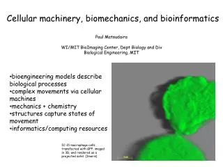
Cellular machinery, biomechanics, and bioinformatics. Paul Matsudaira WI/MIT BioImaging Center, Dept Biology and Div Biological Engineering, MIT. bioengineering models describe biological processes complex movements via cellular machines mechanics + chemistry

E N D
Cellular machinery, biomechanics, and bioinformatics Paul Matsudaira WI/MIT BioImaging Center, Dept Biology and Div Biological Engineering, MIT • bioengineering models describe biological processes • complex movements via cellular machines • mechanics + chemistry • structures capture states of movement • informatics/computing resources IC-21 macrophage cells transfected with GFP, imaged in 3D, and rendered as a projected solid (Imaris)
Quantitative Biology Paradigm courtesy of CSBi
Cell biologists study large, dynamic structures G. Borisy and S. Tatiana
Overlapped structural and cell biology approaches single particle det. dynamic 4D light microscopy cryo electron microscopy NMR, x-rays resolution and size scale 1 nm 1 µm 1 mm protein machines molecules organelles cells
Forces generate movements, machines generate forces zone of adhesion and extenstion fimbrin-EGFP timelapse 30 sec
Changes in structure-assembly, force, chemistry mother adhesion daughter adhesions top side Formation of cell adhesions at the leading edge of a macrophage cell (J. Evans et al J. Cell Biol. in press)
Cell Biology Appetite for More Information • multispectral fluorescence data acquisition • video rate data acquisition • cellular tomography and single particle cryoEM imaging • high content screening • whole proteome studies
High content: Tracking all cell adhesions (J. Evans et al J. Cell Biol. in press)
Dynamics of structure modulated by microtubules 10 µm paclitaxel 10 µm demecolcine (J. Evans et al J. Cell Biol. in press)
Imaging storage/processing requirements object file size genome sequence 12 MB protein structures1 2 GB 2D localization2 6 GB 3D localization3 300 GB 4D localization4 3.6 TB 152kDa (ave. MW) x 110 MW/residues x 10 atoms 2512x512 two channel, 16 bit TIFF 350 image slices/stack 412 images/series
The BioImaging Pipeline acquisition processing analysis management modelling
Podosomes split, merge, and appear de novo dendroid simple polar assembly
Imaging is an informatics science genomics imaging traces counts ATGC voxel sequence image function function
terminal terminal terminal terminal Origin 3400 Origin 2400 4 TB 4 TB WI/MIT BioImaging Net Tape imaging modes confocal microscopy RAID 30TB Cryo-EM 2-photon microscopy IBM Power4 6550
Expansion and management of imaging data multi-spectral (channels) 12 or 16-bit acquisition channel 0 channel 1 channel 2 1+2+3 image size (pixels) channels file size 256 x 256 1 130 KB 3 910 KB 1024x 1024 1 2 MB 3 14 MB
3D/4D image data (stacks) 35KB to 8.2 MB per channel/stack (X:Y:Z or X:Y:T) (x,y,z,t,ch)file size 256 x 256 x 1 x 3 400 KB 256 x 256 x 50 x 3 20 MB 256 x 256 x 50x 20 x 3 400 MB 1024 x 1024 x 50 x 3 800 MB 1024 x 1024 x 50x 20 x 3 16 GB
NMR x-ray light microscopy cryoEM
Image processing (deconvolution) RAM 256 x 256 x 50 x 3 (x,y,z,ch) raw data 12 or 16-bit raw data 32-bit intermediates 32-bit deconvolved data 32-bit 40 MB 80 MB 80 MB 80 MB 80 MB accumulated file size 40 MB 120 MB 200 MB 280 MB 360 MB
Total data accumulation rendering movies raw 0.4 GB post-deconvol 3.6 GB analysis 7.2 GB total 11.2 GB 1 GB 1 GB colocalization object analyses ch1 vs. ch2 ch1 vs. ch3 ch2 vs. ch3 ch1 vs ch2 vs. ch3 1 GB ? 3.2 GB
terminal terminal terminal terminal Current WI (local) imaging net SGI Origin 3400 Zeiss 510 META ftp • stack (512 x 512 x 50 x 12 x 6) 4.8 GB 32 GB RAM 4 TB data acquisition data storage image restoration image analysis batch rendering database management remote visualization rendering batch design
Microtubules at podosomes tubulin actin fimbrin
Dynamic structures from light microscopy assembly of cell adhesions in a live cell ‘finger’ ‘finger’ ‘palm’ ‘palm’ ‘finger-tip’ side/bottom view top view fimbrin-EGFP timelapse 15 sec