Download

1 / 19
190 likes | 343 Views
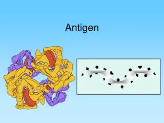
Micro-Fluidic Device for Antigen Discovery. By: Khine Lwin August 23, 2007 Graduate Student: Armando Tovar Professor: Dr. Abraham Lee. Background. Antigen: a foreign molecule that triggers an immune response upon entering the body

E N D
Micro-Fluidic Device for Antigen Discovery By: Khine Lwin August 23, 2007 Graduate Student: Armando Tovar Professor: Dr. Abraham Lee
Background • Antigen: a foreign molecule that triggers an immune response upon entering the body • Antibody: a protein that attaches to antigens, tags them as foreign, and neutralizes them Obtained from Wikipedia.org
Purpose • Create a micro-fluidic device that can detect diseases quickly and efficiently • Faster than ELISA (takes about 24 hours) • Simple design • Diagnostics: • Detect serum concentrations • West Nile, HIV, Smallpox, bioterrorist threats, etc. • Detect food allergens Device consists of: • Ti/Au electrode array • Micro-channel created using polydimethylsiloxane (PDMS) • H3L Proteins immobilized on electrode array Figure 1. Micro-fluidic Device
Photolithography Figure 2. Photolithography Process
Protein Immobilization Surface Printing Tip (SPT) Nano Ware Nano eNabler Printing Process
System Current Amplifier Function Generator DMM Faradic Cage Device Current Amplifier DAQ Computer Flow AC R Equivalent Circuit Figure 5. Flowchart of System
Channel Cs Bulk Solution Rs Double Layer Antibody Diffusion layer CDL Antigen CIL Immobile layer
Double Layer CDouble Layer
Experimental Procedure Figure 2. Flow chart of system Table 1. Protocol for Testing Micro-fluidic Device
Results Figure 6. Signal Response as Flow is Turned On and Off
PDMS • Current Design • 1x .5 mm Electrode Tips • 200 µm PDMS channel • 10 µm gap between the Electrode Tips Figure 4. PDMS Channel Sealed on top of Electrode Tips
Future Work • Improve LabVIEW program • Further research how channel alignment affects impedance • Improve methods for fluorescently tagging antibodies to confirm antigen-antibody binding
Special Thanks • Dr. Abraham Lee • Armando Tovar • BioMint Lab • IM-SURE • National Science Foundation • Said Shokair • Jason Choi & Lillian Shido Any Questions?
LabVIEW Current Program: Prompt user to save file Graph voltage root mean square Extract data and calculate admittance Save data to spreadsheet in Excel Figure 7. User-interface of current LabView program