Download
1 / 12
130 likes | 503 Views
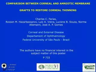
COMPARISON BETWEEN CORNEAL AND AMNIOTIC MEMBRANE GRAFTS TO RESTORE CORNEAL THINNING. Charles C. Farias, Rossen M. Hazarbassanov, Luiz A. Vieira, Luciene B. Souza, Norma Allemann, José A. P. Gomes Corneal and External Disease Departament of Ophthalmology

E N D
COMPARISON BETWEEN CORNEAL AND AMNIOTIC MEMBRANE GRAFTS TO RESTORE CORNEAL THINNING Charles C. Farias, Rossen M. Hazarbassanov, Luiz A. Vieira, Luciene B. Souza, Norma Allemann, José A. P. Gomes Corneal and External Disease Departament of Ophthalmology Federal University of São Paulo - Brazil The authors have no financial interest in the subject matter of this poster P 733
PURPOSE To evaluate the use of preserved corneal and multilayerAM graft for the surgical repair of corneal thinning. PARTICIPANTS • Prospective, comparative, interventional and controlled study • 19 eyes of 19 patients • The mean age was 61,05±SD (45-80) • 09 were female and 10 male • All patients were operated by one surgeon (CCF) • Surgical procedure was randomized: 1. Corneal graft (10 eyes) 2. AM graft(9 eyes)
METHODS • Complete eye examination: BCVA, Biomicroscopy, Tonometry, Fundoscopy, UBM, USG • Surgical Technique: superficial keratectomy, regularity of the thinning edge with trephine, ressection of excessive tissue, pocket formation, preparation of tissue donor, 10-0 nylon suture • Posoperative • Topical prednisolone acetate 1% • Topical ofloxacine 0,3% • All suture were removed within 3 months • Follow up • One day, 7, 15, 30, 90 and 180 days after surgery.
RESULTS Time of epithelization ( 6m) Thickness (6m) AM Cornea Cornea AM AM AM Group Group Mann-Whitney Test, p=0,165 Mann-Whitney Test; p=0,216
RESULTS BCVA(LogMar) 6m AM Mann-Whitney Test; p=0,457
Amniotic membrane graft group DM-Diabetes Mellitus; HBP- high blood pressure; DYSL- Dyslipidemia;
Corneal graft group DM-Diabetes Mellitus; HBP- high blood pressure; DYSL- Dyslipidemia; CARD-Cardiopathies
Pre operative of corneal thinning 7th day pos op of amniotic membrane transplantation 6 months pos op of amniotic membrane transplantation
Pre operative of corneal thinning 7th day pos op of corneal graft 6 months pos op of corneal graft
RESULTS • All eyes that received corneal grafts (10/10) presented stability of the ocular surface with rapid re-epithelialization and restauration of the corneal thickness • Eyes that received the AM grafts also presented stability of the OS with re-epithelialization (8/9), but the transplanted tissue absorbed on average after 30 d of follow up (p<0.05) • 1/9 partial AM re-absortion • 8/9 total AM re-absortion • A persistent epithelial defect was noted in in only two eyes
DISCUSSION • In the present study we observed • a marked reduction in ocular inflammation after AMT • Corneal stroma thickness was restored in 88,88% and 100% of cases after AMT and corneal graft, respectively • rapid epithelial wound healing and long-term stability of the corneal surface
CONCLUSION The results suggest that both AM and corneal grafts are good options to be used for restoring corneal defects with thinning.