Download
1 / 12
280 likes | 3.17k Views
Corneal Hysteresis. Role of Corneal Biomechanics in Refractive Surgery. Luis Rodriguez, MD Anny Villegas, MD Cl ínica de Córnea Centro Médico Docente La Trinidad Caracas, Venezuela.

E N D
Corneal Hysteresis Role of Corneal Biomechanics in Refractive Surgery Luis Rodriguez, MD Anny Villegas, MD Clínica de Córnea Centro Médico Docente La Trinidad Caracas, Venezuela
Corneal Biomechanics is the science that studies the equilibrium and deformation of tissues submitted to any force. This science includes the following extrinsecal factors: Intraocular Pressure, Cilliar Muscle, Extraocular Muscle, Atmospheric Pressure and Eyelids. Intrinsecal factors of Corneal Biomechanics include: Central Pachymetry, Viscosity, Elasticity, Hydration and Regional Pachymetry.
The Ocular Response Analyzer (ORA) is a new instrumentthat measures the corneal biomechanical response (corneal hysteresis,CH). It measures the effect of viscous and elastic qualities with a calibrated air pulse in vivo. The ORA measures Intraocular Pressure (Goldmann). Corneal hysteresis is an indication of viscous damping in thecornea, reflecting the capacity of corneal tissue to absorb anddissipate energy. It is the capacity of tissue to recover its original shape after external force is applied.
The Corneal Resistance Factor, also derived from cornealhysteresis, is an indicator of the overall resistance of the cornea. Corneal-compensated IOP is a pressure measurement thatutilizes the new information provided by the corneal hysteresismeasurement to provide an IOP that is less affected by cornealproperties. The objective of this study is to discuss the Ocular Response Analyzer readings of corneal biomechanics in refractive surgery and corneal pathology. This study began in July 2006 and ended in January 2007.
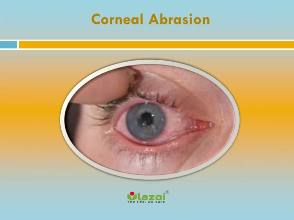
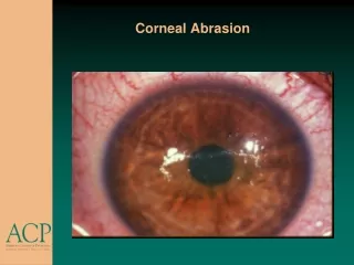
Methods ORA was used to obtain four measurements in each patient, and the mean of these four readings was used in the analysis according to the manufacturer’s instructions. The study included 244 eyes from the Corneal Department of the Centro Medico Docente La Trinidad divided into the following nine categories: Normal cornea, Radial Keratotomy, PostLasik Myopia, PostLasik Ectasia, Keratoconus, Post Intracorneal Segments (Intacs®), Penetrating Keratoplasty, Fuchs’ Dystrophy and Graft rejection.
7,96 – 11,57 20 Normal cornea 10 Radial Keratotomy 3,50 – 5,75 3,42 – 7,43 10 Post-LASIK Ectasia 7,24 – 9,82 34 Post-LASIK Myopia 5,74 – 7,71 70 Keratoconus 70 Post-Intracorneal Segments (INTACS®) 7,09 – 9,97 7,74 – 11,71 20 Penetrating Keratoplasty 6,25 – 7,71 5 Fuch’s Distrophy 5,35 – 7,40 5 Graft Rejection Results Ranges of Corneal Hysteresis (mmHg) 244 Eyes
10 9 8.24 8.93 7.73 7.71 8.62 8 9 8.24 7 6.3 6.95 8 6.47 5.42 6 7 5 6 4 5 Post - LASIK Normal Cornea 3 Post -INTACS 4 Post -LASIK Ectasia 2 Keratoconus 3 1 0 2 1 Normal Cornea 0 Post-LASIK Post-INTACS Keratoconus Post-LASIK Ectasia Frequency of Corneal Hysteresis Frequency of Corneal Resistance Factor (CRF)
Frequency of IOPg and IOPcc values 18 16,36 15,05 16 14,86 14,73 14,49 14,23 14 11,64 11,22 11,54 12 10 9.02 8 6 4 2 0 Normal Cornea Keratoconus Post-LASIK Post- LASIK Ectasia Post-INTACS® IOPg IOP cc
Keratoconus Lasik Ectasia Post Lasik
Discussion Patients with -6.00D (equal to or less), Hysteresis of more than 11mmHg, and normal topography are good candidates for LASIK. Patients with -6.00D or less, suspicious topography and Hysteresis between 8mmHg and 10mmHg are good candidates for PRK. Patients with -6.00D or more, suspicious topography and Hysteresis between 7mmHg and 10mmHg are good candidates for Phakic IOL. Patients, with Keratoconus and Hysteresis of more than 7mmHg are good candidates for intracorneal segments. Patients with Keratoconus and Hysteresis of equal to or less than 5mmHg are candidates for corneal transplant. When Hysteresis is less than 7mmHg PostLasik, patients have Ectasia. Hysteresis is normal in Corneal Transplants, but if the patient has a transplant rejection Hysteresis decreases significantly.
Biomechanical studies must be analyzed individually • Ectasia is a known risk of LASIK and PRK • Hysteresis measures corneal biomechanical properties • Patients who are at risk of developing post-LASIK ectasia should be identified by analyzing Hysteresis • Corneal Hysteresis can guide the physician in the choice of treatment. • ORA helps us understand the IOP in patients with corneal pathologies and surgeries.