Cardiac Tamponade
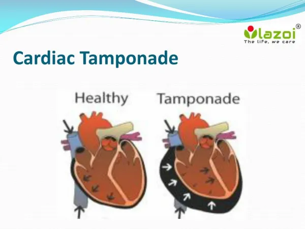
Cardiac Tamponade. Objectives. The learner will be able to: Describe cardiac tamponade and its signs and symptoms. Discuss management strategies for cardiac tamponade. Definition. Pericardial pressure > 30 mm Hg from fluid accumulation (effusion) in the pericardial sac

Cardiac Tamponade
E N D
Presentation Transcript
Objectives The learner will be able to: • Describe cardiac tamponadeand its signs and symptoms. • Discuss management strategies for cardiac tamponade.
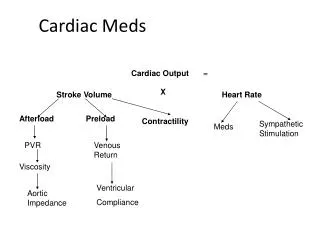
Definition • Pericardial pressure > 30 mm Hg from fluid accumulation (effusion) in the pericardial sac • Pressure on chambers inhibits inflow of blood to the ventricles and reduces cardiac output.
Significance • Tamponade can occur with only 5080 mL of accumulation. • Untreated can lead to: • Cardiovascular collapse • Shock • Death
Disease-Related Incidence • Primary malignancy • Malignant mesothelioma • Histiocytoma • Rhabdomyosarcoma • Angiosarcoma • Pericardial effusions • Present in 5%50% of all malignancies • Thoracic cancers (lung, breast, and lymphoma)
Treatment-Related Incidence • History of radiation to the chest • Chemotherapy • Doxorubicin, daunorubicin, and cyclophosphamide • Biotherapy • Interferon, interleukin, and colony-stimulating factors
Early Symptoms (Effusion) • Asymptomatic during “effusion” period • Often mimic heart failure • Jugular venous distention (JVD) • Peripheral edema • Hepatomegaly and abdominal distention • Increased diastolic pressure • Tachycardia • Fatigue, dyspnea, and orthopnea
Late Symptoms • Dull chest pain/heaviness • Increasing dyspnea and “air hunger” • Tripod positioning • Nonproductive cough • Anxiety, agitation, and mental status changes • Cold sweats or confusion • Hiccoughs, dysphagia, or hoarse voice
Physical Assessment • JVD (nonpulsating) • Weak pulses and tachycardia • Muffled heart sounds; possible friction rub • Downward, left shift of PMI • Hypotension and narrow pulse pressure • Decreased urine output (oliguria) • Pulsus paradoxus (late sign)
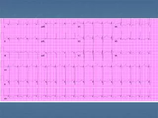
Diagnostics • Chest x-ray • EKG changes • CT or MRI scans • Transesophogeal echocardiogram • Pericardiocentesis
Treatment • Immediate removal of fluid • Pericardiocentesis • Control reaccumulation of fluid. • Permanent defect in pericardium to drain • Sclerosis of mesothelium with irritants given via catheter to prevent fluid reaccumulation
Pericardiocentesis • Complications (occur in 10%25%) • Puncture of cardiac muscle or artery • Air emboli • Dysrhythmia • Infection/abscess • Vagal response and bradycardia
Additional Therapies • Radiation therapy • Chemotherapy • Glucocorticoids • IV fluids • Diuretics
Nursing Management • Early identification of risk and manifestation • Assist with positioning and activities. • Oxygen • Manage pain. • Manage anxiety. • Ongoing regular assessments • Prepare for procedures.
References Braunwald, E. (2001). Pericardial disease. In E. Braunwald, A.S. Fauci, D.L. Kasper, S.L. Hauser, D.L. Longo, & J.L. Jameson (Eds.), Harrison’s principles of internal medicine (5th ed., pp. 13651372). New York, NY: McGraw-Hill. Flounders, J.A. (2003). Cardiovascular emergencies: Pericardial effusion and cardiac tamponade [Online exclusive]. Oncology Nursing Forum, 30, E48E55. Grannis, F.W., Wagman, L.D., Lai, L., & Curcio, L.D. (2002). Fluid complications. In R. Pazdur, L.R. Coia, W.J. Hoskins, & L.D. Wagman (Eds.), Cancer management: A multi-disciplinary approach (pp. 943958). Melville, NY: PRR. Hawley, J., Dreher, H.M., & Vasso, M. (2003). Under pressure: Treating cardiac tamponade. Nursing Management,34(2), 44D, 44F, 44H. Humphreys, M. (2003). Conditions affecting the pericardium. Connect, The World of Critical Care Nursing, 2, 8084.
References (cont.) Hunter, J.C. (2005). Structural emergencies. In J.K. Itano & K.N. Taoka (Eds.), Core curriculum for oncology nurses (4th ed., pp. 442439). St. Louis, MO: Elsevier Saunders. Kaplow, R. (2005). Cardiac tamponade. In C. Yarbro, M.H. Frogge, & M. Goodman (Eds.), Cancer nursing: Principles and practice (6th ed., pp. 837886). Sudbury, MA: Jones and Bartlett. Keefe, D.L. (2000). Cardiovascular emergencies in the cancer patient. Seminars in Oncology, 27, 244255. Myers, J.S. (2001). Oncologic complications. In S.E. Otto (Ed.), Oncology nursing (4th ed., pp. 498581). St. Louis, MO: Mosby. Read, W., & Denes, A. (2002). Oncologic emergencies. In R. Govindan (Ed.), Washington manual of oncology (pp. 465466). Philadelphia, PA: Lippincott Williams and Wilkins.
References (cont.) Shabetai, R. (2004). Pericardial effusion: Haemodynamic spectrum. Heart, 90, 255256. Shatzer, M., & Castor, A. (2004). How transthoracic echocardiography detects cardiac tamponade. Nursing, 34(3), 7374. Shelton, B.K. (2000). Pericarditis/pericardial effusion/cardiac tamponade. In D. Camp-Sorrell & R.A. Hawkins (Eds.), Clinical manual for the oncology advanced practice nurse (pp. 307326). Pittsburgh, PA: Oncology Nursing Society. Story, K.T. (2006). Cardiac tamponade. In M. Kaplan (Ed.), Understanding and managing oncologic emergencies: A resource for nurses (pp. 129). Pittsburgh, PA: Oncology Nursing Society.