Download
1 / 41
420 likes | 562 Views
The changing face of cancer diagnosis. George Vassiliou November 2011. Overview. Today’s cancer diagnostic lab The era of cancer genomics Novel diagnostic applications Introducing genomics to cancer diagnosis. Overview. Today’s cancer diagnostic lab The era of cancer genomics

E N D
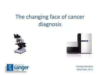
The changing face of cancer diagnosis George Vassiliou November 2011
Overview • Today’s cancer diagnostic lab • The era of cancer genomics • Novel diagnostic applications • Introducing genomics to cancer diagnosis
Overview • Today’s cancer diagnostic lab • The era of cancer genomics • Novel diagnostic applications • Introducing genomics to cancer diagnosis
The light microscope remains the central cancer diagnostic tool for 400 years Zacharias and Hans Jansen (ca 1595) Modern microscope (ca 1995)
Today’s cancer diagnostic lab Cellular Phenotyping Microscopy (histology/cytology) Immunohistochemistry Flow Cytometry Genetic tests Cytogenetics Molecular Genetics Genotyping for specific mutations (PCR/RT-PCR) Minimal Residual Disease monitoring (CGH and SNP/LOH genotyping) (Gene Expression Profiling)
Haemato-oncology lab Sample Microscopy Immunophenotyping Cytogenetics Molecular Genetics Cytology Histology Immunohistochemistry Diagnostic panels Karyotyping FISH Mutational screening RT-PCR qPCR (MRD) Integrated report MICROSCOPY: >80% undifferentiated blasts Morphology of acute lymphoblastic leukaemia Eosinophils, basophils and small megakaryocytes suggest blast phase of chronic myeloid leukaemia IMMUNOPHENOTYPE: Blast cells are CD10, CD19, CD79a, CD34, HLA-DR, TdT positive. Weak CD13. They do not express CD33 or myeloperoxidase. DNA index is 1.0 Phenotype of B lymphoblastic leukaemia or B lymphoblastic transformation of CML CYTOGENETICS: Karyotype: 46,XY,t(9;22)(q34;q11) in 10 of 10 metaphases FISH: BCR/ABL 92% positive MOLECULAR GENETICS: BCR-ABL fusion transcript type: p210 e13a2 by RT-PCR OVERALL CONCLUSION: B lymphoblastic blast crisis of chronic myeloid leukaemia
Diagnostic gene expression profiling n=241 MammaPrint – 70 gene signature (NKI) Lymph node positive breast cancer
Overview • Today’s cancer diagnostic lab • The era of cancer genomics • Novel diagnostic applications • Introducing genomics to cancer diagnosis
Advances in DNA sequencing technologies Ion Torrent 1016 1014 1012 Output kbp / run 1010 Capillary (Sanger) Sequencing Next Generation Sequencing (NGS) 108 106 104 102 ABI capillary 454 pyroseq Solexa/ Illumina ABI SOLID ABI SOLID 3.0 Illumina HiSeq Roche/454 Titanium Technologies 2003 2004 2005 2001 2002 2006 2007 2008 2009 2010 2011
Sanger Institute Total yield by week (Gigabases) 2008 2009 2010 2011
How Fast is That? 6000 Gb per week (6 Tb) = 10,000,000 bases per second ½ hour per 6Gb (= 1x Human Genome)
Genome Sequencing Sanger (capillary) sequencing Next generation sequencing Cancer Genomics Melanoma Small-cell lung Breast Myeloma CLL Mouse AML AML Lung (NSS) Hepatocellular 2015 ? ~1day ?? $100 2000 ~10 years ~$ 3.5 billion 2005 ~3 years ~$ 20million 2010 ~1month $9,500 (Illumina) 2008 ~4 months ~$ 1.5million
Analysing cancer genomes From Ding et al, Hum Mol Gen, 2010
Genomic Circos Plot Genomic coordinates Deletions/Insertions Substitution density (het) Substitution density (homo) Coding Substitutions Silent Missense Nonsense Splice site Copy number Regions of LOH Structural rearrangements Intrachromosomal Interchromosomal Circos Plot from Pleasance et al, Nature 2010
This decade • 1000sof individual cancer genomes • 100sof recurrent mutations • Aetiological links • Clinico-pathological correlates • Delineation of effects of many mutations • Development of new therapies > Increasing use of genomics in cancer diagnosis, prognosis & therapy
Overview • Today’s cancer diagnostic lab • The era of cancer genomics • Novel diagnostic applications • Introducing genomics to cancer diagnosis
The clinical process in oncology Pre-clinical phase Presentation Relapse Diagnosis Follow-up Treatment Assessment of response
New diagnostic applications • Whole genome sequencing (~6Gb) • Exome sequencing (~60Mb) • Selected gene/exon DNA sequencing • Residual disease monitoring (plasma DNA)
Whole genome sequencing B A D C
Diagnostic whole genome sequencing Constitutional genome Cancer genome Compare Inherited mutations & polymorphisms Substitutions Indels Copy number changes Translocations Somatic mutations Subclonal heterogeneity Other diagnostic data Clinical Report
Exome sequencing: target enrichment Chr1 NRAS “Baits” or PCR amplicons regions covered
Diagnostic whole exome sequencing Constitutional exome Cancer exome Compare Inherited mutations & polymorphisms Substitutions Indels Copy number changes Translocations Somatic mutations Subclonal heterogeneity Other diagnostic data Clinical Report
Selective sequencingexample: an AML toolkit • ~20 genes known to be recurrently mutated • Prognostic/treatment implications known for some • Target enrichment by “pull down” Can detect: Sequence changes Copy number (UPD/LOH)
Plasma DNA Normal tissues Cancer DNA with tumour-specific mutation Slide courtesy of Dr Peter Campbell
Tumour-specific rearrangements Chr10 Chr20 1st round PCR Nested real-time PCR Individual Breast Cancer Genome
Relapsing breast cancer Non-rearranged genomic region Tumour-specific rearrangement Undiluted patient plasma 1:10 1:100 1 1:1000 1:10,000 1:100,000 0.5 Normal 1 Water 0.5 0.1 Intensity Intensity 0.05 0.1 0.05 0.01 0.01 0 5 10 15 20 25 30 35 40 0 5 10 15 20 25 30 35 40 Cycles of real-time PCR Cycles of real-time PCR Slide courtesy of Dr Peter Campbell
Serial measurements CT scan: Localised deposits around T9-10 CT scan: Widespread soft-tissue metastases Chemotherapy: 150 First-line chemotherapy Second-line Paclitaxel 125 100 Estimated tumour DNA / mL serum (pg) 75 Rearrangement 1 Rearrangement 2 50 25 Detectable at limit of sensitivity Undetectable 5 6 7 8 9 10 11 12 13 14 15 16 17 Months after diagnosis Slide courtesy of Dr Peter Campbell
Other applications of NGS • Multiple biomarker MRD • Methylomics • Transcriptomics (RNAseq) • Cancer screening / Biomarker assays
Overview • Today’s cancer diagnostic lab • The era of cancer genomics • Novel diagnostic applications • Introducing genomics to cancer diagnosis
Hurdles to the introduction of diagnostic cancer genome sequencing • Clinical relevance/utility Evolving • Personal genomes/Ethics Being tested Clinic • Sample choice/compatibility FFPE, other • Cost $10,000/genome • Sample to sequence delay 8-10 days • Mutation calling Specificity / Sensitivity Technology • Sequencing equipment Choice/Cost • New personnel Bioinformaticians • Computer storage Petabytes (1015) • Education/training Pathologists, Clinicians Laboratory
Data storage & analysis Genome Campus Research Support Facility Data Centre Sulston Building Morgan Building European Bioinformatics Institute
Training of pathologists • Core training in genomics • New sub-specialty e.g. Molecular Pathology? • Impact on other aspects of training • How will the training be delivered? • Training/role of laboratory scientists • Keeping control of the agenda
Diagnostic reporting of genomic data • Communicating the cancer genome to the clinician • Diagnosis • Recurrently mutated genes • Non-recurrent/private mutations / pathways • Prognostic relevance • Therapeutic relevance • Pharmacogenomics • Constitutional genome • Mutational signatures • Summary / Imagery
Implications for cancer classification Cellular origin Morphology Differentiation/grading Mutations: Diagnosis Prognosis Treatment Unified Classification?
Acute Myeloid Leukaemia – a paradigm of evolving classification
Summary • The advent of cancer genomics is changing cancer medicine • Changes will transform cancer diagnosis and the role of pathologists • Pathologists need to understand what is coming in order to lead and formulate the future for cancer diagnosis