Download
1 / 25
260 likes | 583 Views
Facial Soft Tissue Infections. Heather Patterson PGY-4 November 13, 2008. Objectives. By the end of this session the learner will be able to outline clinical features, management strategies, and complication of facial infections including: Cellulitis Erysipelas Orbital Cellulitis

E N D
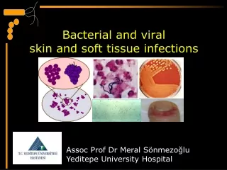
Facial Soft Tissue Infections Heather Patterson PGY-4 November 13, 2008
Objectives • By the end of this session the learner will be able to outline clinical features, management strategies, and complication of facial infections including: • Cellulitis • Erysipelas • Orbital Cellulitis • Periorbital Cellulitis
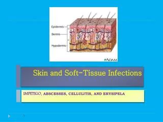
Cellulitis • Def’n: • Soft tissue infection of the skin and subcutaneous tissue • Risk Factors: • Skin trauma • Lymphatic or venous stasis • FB • Immunosuppression
Cellulitis • Clinical Features: • Skin: • Red, swollen, warm, painful • Blanching • +/- lymphadenopathy • Vitals • +/- tachycardia, otherwise normal vitals • Labs: • Minimal change to WBC • Pertinent negatives • Fever uncommon • No crepitus or bullae
Cellulitis • Ddx: • Orbital/preorbital • Erysipelas • Impetigo • Folliculitis • FB • Fascitis • Myositis • Post surgical healing • Burn
Cellulitis • Bugs and Drugs: • Staph and Strep • Gram negative • MRSA
Erysipelas • What is erysipelas? • What does it look like? • Who get erysipelas? • How do we treat it?
Erysipelas • What is erysipelas? • Superficial cellulitis involving dermis, lymphatics, and most of the superficial subcutaneous tissue
Erysipelas • What does it look like? • Sharply demarcated border +/- vessicles at margin • Raised • Dark erythema • Indurated • Other features: • Toxic appearing pt with prodrome of fever, chills, malaise,vomiting • Rapid spread, very painful, itchy, burning • Prominent lymphadenopathy
Erysipelas • Who gets this? • Young or >50y • Risk factors: • EtOH abuse, venous stasis, DM, nephrotic syndrome • Associated with small breaks in the skin, post operative infections
Erysipelas • How do we treat it? • MCC Group A Strep • Pen G or erythromycin • Cephalosporins, macrolides, fluoroquinolones for more severe cases
Orbital and Periorbital Cellulitis • Anatomic differences • Epidemiology • Pathophysiology • Clinical Features • Management • Complications
Orbital and Periorbital Cellulitis • What is the difference in the location of infection? • Periorbital - preseptal • Orbital - posterior to the orbital septum
Orbital and Periorbital Cellulitis • What is the population at risk? (i.e. epidemiology) • Children / adolescents + older pts • Pathophysiology: • Extension from surrounding infections: • Coexisting sinusitis in 80% • Dental infections • Direct innoculation: • Facial trauma • Hematogenous spread • Vascular lesions, chemical agents
Orbital and Periorbital Cellulitis • What are the common bugs involved? • Staph and strep • Hflu (if unimmunized) • Differentiate between the clinical presentation of the 2 entities: • Skin findings • Occular findings
Orbital and Periorbital Cellulitis • What are the complications associated with orbital and periorbital cellulitis? • Orbital cellulitis: • Orbital abscess • Subperiostal abscess • Loss of vision • Optic neuritis • Retinal vein thrombosis • CNS extension • Meningitis, abscess • Cavernous sinus thrombosis
Orbital and Periorbital Cellulitis • What are the management strategies? • Orbital • Rapid dx - CT • Ophtho consult • Abx: amp/gent/flagyl or Clinda/gent or Ceftriaxone/flagyl • What about lateral canthotomy? Indications? Procedure? • Periorbital • R/O orbital ceullulitis • Abx: Cefuroxime x 2/7 and then po • Admit if unwell or indicated by social situation
Lateral Canthotomy • Goals: • Rapidly decrease IOP • Reinstitute retinal artery blood flow • Steps • Simple, rapid saline cleaning of lids • Anesthetize with 1-2% lidocaine with epi • Crush lateral canthus 1-2min with hemostat • Incise lateral canthus with iris scissors • Incision extends toward orbital rim • Identify superior and inferior crus of lateral canthal tendon • Release inferior canthal tendon
Cavernous Sinus Thrombosis • Clinical Presentation • Headache, fever, malaise • Face: • Midface infection or sinusitis • Periorbital edema, proptosis, ptosis, orbital pain, chemosis • Occular exam • Sluggish pupillary response, decreased acuity, papilledema, • CNS: • CN findings (CN VI first) EOM • Mental status changes, confusion, drowsiness
Cavernous Sinus Thrombosis • Management: • Early diagnosis • Early Abx • Anticoagulation? • Bhatia et al 2002 • Steroids • Surgery is NOT indicated