Compartment Syndrome
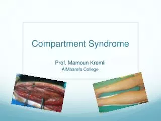
Compartment Syndrome. Kendra Morrison, DO EM PGY 4 SJHC. Tissue pressure rises confined space Tissue hypoxia damage to compartment structures (nerves,arteries,veins,muscles) Irreversible damage (Volkmann’s contracture) Elevated compartment pressures

Compartment Syndrome
E N D
Presentation Transcript
Compartment Syndrome Kendra Morrison, DO EM PGY 4 SJHC
Tissue pressure rises confined space • Tissue hypoxia damage to compartment structures (nerves,arteries,veins,muscles) • Irreversible damage (Volkmann’s contracture) • Elevated compartment pressures • Extrinsic forces (cast too tight) • Intrinsic forces (increase in volume of contents in compartment like hematoma or edema)
Table 278-1 Common Causes of Compartment Syndrome Orthopedic Tibial fractures (especially if it is a closed injury) Forearm fractures (closed) Vascular Ischemic-reperfusion injury (can occur after closed reduction of fracture or dislocation) Hemorrhage (as in knee dislocation and popliteal aa. Injury) Iatrogenic Vascular puncture in anticoagulated patients Intravenous/intra-arterial drug injection Constructive casts Soft tissue injury Prolonged limb compression Crush injury Burns
Normal tissue perfusion = diff. b/w arterial and venous pressures at capillary level • Compartment pressures increase tissue perfusion compromised by loss of vasomotor tone at arterioles or collapse of thin-walled veins • Muscles become ischemic and inflammatory response initiated • Histamine release • Dilation of capillary beds • Increase in capillary permeability
Normal pressure < 10 mmHg • Capillary blood flow compromised at > 20 mmHg • This will cause pain & is an early finding (pt will poorly localize the pain) • Ischemic necrosis of muscles/nerves > 30-40 mmHg • Nerve more sensitive than muscle (“paresthesias”) • If distal pulses absent, then muscle necrosis has occurred (“pulselessness”)(too late!!) • These muscles hurt and will exacerbate pain with active or passivemotion of extremity • Hence, pain out of proportion to exam (pt will describe as deep & unremitting) • If prolonged high pressures, then paralysis of mm. will eventually occur • Compartment will be tense compared to other side
Compartments • Upper arm (uncommon to occur here) • Anterior • Biceps, brachialis mm.; ulnar, median, radial nn. • Posterior • Triceps mm. • Forearm (compartments separated by interosseous membrane) • Volar • Wrist & finger flexors; radial, ulnar aa.; median, ulnar nn. • Dorsal • Wrist & finger extensors; radial nn.
Hand (4 compartments) • 1. Thenar: intrinsic mm. of thumb • 2. Hypothenar: intrinsic mm. of fingers • 3. Central: ulnar aa. • 4. Interossei: each finger has own
Gluteal (3 compartments) • 1. Tensor mm. of fascia lata • 2. Gluteus medius & minimus • 3. Gluteus maximus; sciatic nn. • Thigh • Anterior • vastus lateralis, vastus intermedius & vastus medialis mm.; sartorius & rectus femoris mm.; femoral aa. & nn. • Medial • Adductor longus, adductor brevis, adductor magnus, gracilis mm. • Posterior • Semimembranosus, semitendinosus, biceps femoris mm.; Sciatic nn.
Leg (4 compartments) • Anterior • Extensor mm. of toes; ant. tibial aa.; deep peroneal nn. (sensory to web spaces of 1st & 2nd toes) • most susceptible to compartment syndrome • Lateral • Fibula; intermuscular septum; peroneal mm. (evert foot); superficial peroneal nn. (innervates peroneal mm. and sensory to lateral dorsal foot) • Posterior • Superficial: gastrocnemius & soleus mm. • Deep: flexor mm. of foot; posterior tibial aa.; tibial & peroneal nn.(sensory to heel)
Pressure Measurement • Stic catheter (Stryker) • Fluid filled syringe on a side-ported needle • Insert into suspected compartment • Inject 3cc of saline • Calibrate to normal compartment pressure (O mmHg) • Highest presssures within a compartment are w/in 5cm of fx site
Treatment • ED FACIOTOMY • LEAVE WOUND OPEN AND PACK WITH WET STERILE GAUZE • CALL ORTHOPEDICS!!