Download
1 / 30
510 likes | 1.9k Views
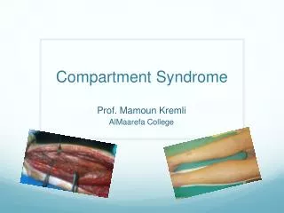
Compartment Syndrome. Prof. Mamoun Kremli AlMaarefa College. Pathophysiology. Increasing volume in a closed compartment P ressure increased in compartment Decreasing arteriovenous difference Hypoxia : Muscle necrosis. Pathophysiology. > 30 mmHg. N=0-4 mmHg. Compartment pressure.

E N D
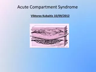
Compartment Syndrome Prof. Mamoun Kremli AlMaarefa College
Pathophysiology • Increasingvolume in a closedcompartment • Pressureincreasedin compartment • Decreasingarteriovenousdifference • Hypoxia : Musclenecrosis
Pathophysiology > 30 mmHg N=0-4 mmHg Compartment pressure Venous outflow Venous pressure Gradient A.V pressure Arterial perfusion Capillary permeability Ischemia, tissue necrosis, edema
Pathophysiology • Increased compartment pressure: • ICP >30mm Hg (>40mm Hg) • Delta Pressure: Pdiast- Pcomp< 30 mm Hg
Causes • Fractures • Bleeding in closedcompartment • Soft tissuetrauma • Bleedingandedema in closedcompartment • Surgery • Post osteotomy (Tibia / Forearm) • Circumfrentialdressings • Does not allowswellingofskin
Clinical Picture – 5Ps • Pain: • Pain out of proportion of expectation • Increased pressure / burst sensation • Pain with passive motion / stretch • Paresthesia • Paralysis • Pallor • Pulselessness TREAT too late, >8h
Clinical Picture - Look • Shiny skin • Pallor / or Dusky skin • Swelling of compartment
Clinical Picture - Look • Shiny skin • Pallor / or Dusky skin • Increased volume • Blisters • Clear fluid • Dusky • Bloody -worst
Clinical Picture - Feel • Feels tense • Parasthesia • Pulse ?
Clinical Picture - Move • Pain on passive stretch • Passive dorsiflexion of ankle (leg) • Passive dorsiflexion of wrist (forearm)
Diagnosis • Diagnosis is clinical: • Unrelenting, burstingpain • Unrelifedbyanalgesia • Swollencompartment • Pain on passive stretching • Sensorydeficit? • Pulses always palpable • Open fractures DO NOT necessarily decompress an elevated compartment pressure
Diagnosis • Compartmentpressuremeasurement: • NOT a substitute for clinical diagnosis • Invaluable in unconscious or anesthetized patients
Measuring compart. pressure • When is pressure measurement needed? • Measure pressure only if: • Clinical picture equivocal • Altered consciousness • Multiple injuries • Epidural anesthesia • Concomitant nerve injury • Children
Treatment • Medical • Surgical
Medical Management • ABC’s. • Correct hypotension • Remove circumferential bandages & cast • Limb at level of the heart • more elevation reduces the arterial inflow • Supplemental oxygen administration
Medical Management • With tight cast, compartmental pressure falls: • 30% when cast is split on one side • 65% when cast is split Bilaterally • 75% with Splitting the inside padding • 85 – 90% complete removal of cast
Surgical Management • Should not be delayed • Fasciotomy • Skin and All compartments
Fasciotomy • Indications: • High suspicion • Unequivocal clinical findings • Significant tissue injury • Delta pressure (DBP - compartment P.) < 25 mm Hg. • Compartment pressure > 30mm Hg. • S&S not resolved after 30-60min of appropriate precautions • Prophylactic with major corrective osteotomy of the leg & forearm • High risk patients
High Risk Patients • Clinical picture equivocal • Altered consciousness • Multiple injuries • Epidural anesthesia • Concomitant nerve injury • Children
Fasciotomy Principles • Long extensile incisions • Release all compartments • Debride necrotic muscles (4C’s) • Preserve neurovascular structures • Never close fascia • Keep wound open • Repeated looks x48h, as needed • Coverage within 7-10 days (usually within 3-5 d)
Fasciotomy Principles • Wound closure: • Bulky dressing with a splint • “Boot lace” vessel loop closure • “V.A.C” dressing (Vacuum Assisted Closure) • Later skin graft / flap: • Usually skin graft • Flap coverage needed if nerves, vessels, or bone exposed
Compartment Syndrome • Evaluation ofmuscleviability (4Cs): • Color • Consistency • Contractility • Capacity to bleed
Treatment - early • Color red • Consistency normal • Capable of bleeding • Contracts when pinched ✓
Treatment – late • Color dark • Consistency abnormal • Not bleeding • No contractions when pinched ✗
Contraindication to fasciotomy • Confirmed acute compartment syndrome diagnosis for > 48 hours • damage cannot be reversed and • significant infection rate when dead tissue exposed • Already dead muscles, as in crush injuries
Complications of untreated C.S. • Volckmann’s contracture • Muscle weakness • Sensory loss • Chronic pain • Amputation
Summary • Compartment syndrome is a clinical diagnosis • Should not be missed - Disaster • Requires urgent treatment • “Time” is the most important factor to avoid irreversible complications • Do NOT apply circumferential dressings