Download

1 / 29
300 likes | 521 Views
THE GENERA CHLAMYDIA and CHLAMYDOPHILA. Characteristics. Rod-shaped or cocciod Obligate intracellular parasites Aerobic Gram negative but difficult to stain Cell w all – lipopolysaccharides form the outer membrane, not peptidoglycan. Forms elementary and reticulate bodies

E N D
Characteristics • Rod-shaped or cocciod • Obligate intracellular parasites • Aerobic • Gram negative but difficult to stain • Cell wall – lipopolysaccharides form the outer membrane, not peptidoglycan. • Forms elementary and reticulate bodies • Non-motile • 37 C, mesophile
ThefamilyChlamydiaceaeconsistsoftwogeneraChlamydiaandChlamydophilawiththree species that cause humandisease: • Chlamydia trachomatis, which can cause urogenital infections, trachoma, conjunctivitis, pneumonia and lymphogranuloma venereum (LGV). • Chlamydophila pneumoniae, which can cause bronchitis, sinusitis, pneumonia and possibly atherosclerosis. • Chlamydophila psittaci, which can cause pneumonia (psittacosis).
Chlamydia are small obligate intracellular parasites and were once considered to be viruses. However, they contain DNA, RNA and ribosomes and make their own proteins and nucleic acids and are now considered to be true bacteria. • They possess an inner and outer membrane similar gram-negative bacteria and a lipopolysaccharide but do not have a peptidoglycan layer. • Although they synthesize most of their metabolic intermediates, they are unable to make their own ATP and thus are energy parasites.
They have a unique growth cycle and their cell wall lack peptidoglycan. • They are currently classified with the gramnegative bacteria within the kingdom Procaryotae.
The chlamydiae stain poorly with the Gram stain, but readily by the Giemsa, Castaneda, Gimenez or Macchiavello methods. • They have a double stranded DNA genome, procaryotic type RNA and synthesize their own proteins during their developmental cycle in membrane-bounded vacuoles in the cytoplasm of host cells. • However, their synthetic processes are dependent on energy (ATP) and metabolites from the host cell pool.
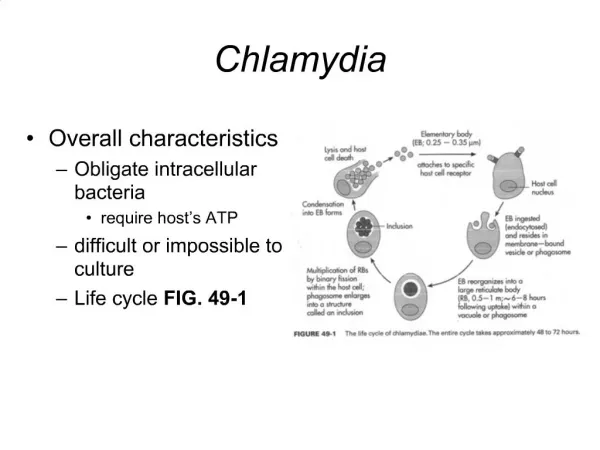
Thechlamydiae are verysmallbacteriawhich are obligateintracellularparasites. Theyhave a more complicatedlifecyclethan free-livingbacteriabecausetheycanexist in twodifferentforms: • the elementary body (EB) is adapted for extracellular survival and for initiation of infection, • the reticulate body (RB) for intracellular multiplication.
Two forms of the chlamydiae are seen in infected cells. • The elementary body (300-400 nm in diameter) is the infectious form. • The initial or reticulate body (800-1200 nm in diameter), with a higher content of RNA, is the metabolically active, non-infectious, fragile form into which the elementary form develops during the multiplication cycle. The reticulate body undergoes a series of divisions by binary fission yielding progeny that are smaller. This culminates in condensation of internal elements, formation of elementary bodies, and their release from the host cell by a phenomenon similar to exocytosis.
Elementary bodies (EB)EBs are the small infectious form of the chlamydia. They possess a rigid outer membrane that is extensively cross-linked by disulfide bonds. Because of their rigid outer membrane the elementary bodies are resistant to harsh environmental conditions encountered when the chlamydia are outside of their eukaryotic host cells. The elementary bodies bind to receptors on host cells and initiate infection. Most chlamydia infect columnar epithelial cells but some can also infect macrophages. • Reticulate bodies (RB) RBs are the non-infectious intracellular from of the chlamydia. They are the metabolically active replicating form. They possess a fragile membrane lacking the extensive disulfide bonds characteristic of the EB.
The rigidity of the cell wall of elementary bodies is facilitated and maintained by extensive disulphide cross-linking of the major outer membrane protein, which is rich in cysteine. • There is a major heat-stable complement fixing, genus-specific antigen, extractable from the microorganism with organic solvens, e.g. ether. This is composed of typical lipopolysaccharide (LPS) components. • Chlamydial LPS shares at two antigenic determinants with the LPS of certain gramnegative bacteria, e.g. Acinetobacter calcoaceticus.
Chlamydiae are sensitive to some antibiotics, notably tetracyclines, macrolides and fluoroquinolones. • Chlamydia trachomatis is sensitive to sulphonamides, but Chlamydophila psittaci, with a few exceptions, is not.
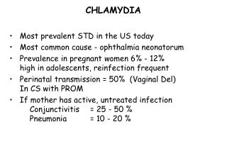
Thechlamydiae cause a widerangeofhumandiseases • The species (Chlamydia trachomatis ) can be subdivided into different serotypes (also known as serovars) and these have been shown to be linked characteristically with different infections. • The majority of infections are genital and are acquired during sexual intercourse. • Asymptomatic infection is common, especially in women.
Ocular infections in adults are probably acquired by auto - inoculation from infected genitalia or by ocular - genital contact. Ocular infections in neonates are acquired during passage through an infected maternal birth canal, and the infant is also at risk of developing Chlamydia trachomatis pneumonia. • Chlamydia trachomatis causes ocular, respiratory, genital tract and probably aural infections, lymphogranuloma venereum (LGV), and some cases of endocarditis and perihepatitis. • Ocular infection take the form of inclusion conjuctivitis in adults or neonates, or trachoma. Genital infections include non-gonococcal or post-gonococcal urethritis, clinical or subclinical cervicitis, epididymitis and salpingitis. A chronic pneumonitis in the newborn has been described.
Chlamydiatrachomatis – biovarsandserovars • C. trachomatis is the causative agent of trachoma, oculogenital disease, infant pneumonia and lymphogranuloma venereum (LGV). • Biovars - C. trachomatis has a limited host range and only infects human epithelial cells (one strain can infect mice). The species is divided into three biovars (biological variants): LGV, trachoma, and mouse pneumonitis. • Serovars - The human biovars have been further subdivided in to several serovars (serological variants; equivalent to serotypes) that differ in their major outer membrane proteins and which are associated with different diseases.
WithinChlamydiatrachomatisthere are threebiovars: • Biovar I with 3 serovars - L1, L2 and L3 which causes Lymphogranuloma venereum (LGV). • Biovar II with 12 serovars A-K, which causes ocular, genital and associated infections. • Biovar III vhich comprises the etiological agent of mouse pneumonitis, the only animal pathogen classified within this species.
Lymphogranuloma venereum is a serious disease which is common in Africa, Asia and South America and occurs sporadically in Europe, Australia and North America. • The prevalence appears to be higher in males than females, probably because symptomatic infection is more common in man.
Treatment • Treatment for chlamydial infection is with tetracycline, macrolides or fluoroquinolones. • It is important to remember that these microorganisms are not susceptible to the beta-lactam antibiotics which are the drugs of choice for treatment of gonorrhoea and syphilis. • Vaccines are of little value and are not used. • Treatment coupled with improved sanitation to prevent reinfection is the best way to control infection. • Safe sexual practices and prompt treatment of symptomatic patients and their sexual partners can prevent genital infections.
Pathogenesisandimmunity • C. trachomatis infects non-ciliated columnar epithelial cells. • The microorganisms stimulate the infiltration of polymorphonuclear cells and lymphocytes which leads to lymphoid follicle formation and fibrotic changes. • The clinical manifestations result from destruction of the cells and the host inflammatory response. • Infection does not stimulate long lasting immunity and reinfection results in a inflammatory response and subsequent tissue damage.
Clinicalsyndromes • Trachoma • Chronic infection or repeated reinfection with C. trachomatis (biovar: trachoma) results in inflammation and follicle formation involving the entire conjunctiva. Scarring of the conjunctiva causes turning in of the eyelids and eventual scarring, ulceration and blood vessel formation in the cornea, resulting in blindness. • Inclusion conjunctivitis • Inclusion conjunctivitis is caused by C. trachomatis (biovar: trachoma) associated with genital infections (serovars D-K). The infection is characterized by a mucopurulent discharge, corneal infiltrates and occasional corneal vascularization. In chronic cases corneal scarring may occur. In neonates infection results from passage through an infected birth canal and becomes apparent after 5-12 days. Ear infection and rhinitis can accompany the ocular disease.
Clinicalsyndromes • Infant pneumonia • Infants infected with C. trachomatis (biovar: trachoma; serovars: D - K) at birth can develop pneumonia. The children develop symptoms of wheezing and cough but not fever. The disease is often preceded by neonatal conjunctivitis. • Ocular lymphogranuloma venereum • Infection with the LGV serovars of C. trachomatis (biovar: LGV) can lead to oculoglandular conjunctivitis. In addition to the conjunctivitis, patients also have an associated lymphadenopathy.
Clinicalsyndromes • Urogenital infections • In females the infection is usually (80%) asymptomatic but symptoms can include cervicitis, urethritis, and salpingitis. Premature delivery and an increased rate of ectopic pregnancy due to salpingitis can occur. In males, the infection is usually (75%) symptomatic. • Reiter's syndrome • Reiter's syndrome is a triad of symptoms that include conjunctivitis, polyarthritis and genital inflammation. • Lymphogranuloma venereum (C. trachomatis biovar: LGV) • The primary lesion of LGV is a small painless and inconspicuous vesicular lesion that appears at the site of infection, often the penis or vagina. The patient may also experience fever, headache and myalgia. The second stage of the disease presents as a marked inflammation of the draining lymph nodes.
Microbiologydiagnosis • Because chlamydiae are obligate intracellular parasites, isolation must be performed in cell cultures. It is used tissue culture McCoy. • After 48-72 hours Chlamydia trachomatis forms characteristic cytoplasmic inclusions which stain with iodine (because they contain glycogen), or can be visualized by immunofluorescent stains. • Chlamydia trachomatis can be detected directly in smears of clinical specimens made on microscope slides, stained with fluorescein - conjugated monoclonal antibodies and viewed by UV microscopy - the direct fluorescent antibody test. Results can be obtained within a few hours. • Chlamydial antigens can also be detected in specimens using an enzyme-linked immunosorbent assay (ELISA).
Chlamydophilapneumoniae • Chlamydophila pneumoniae is a etiologic agent of respiratory tract infection, mainly pneumonia.
Chlamydophilapneumoniae • C. pneumoniae is the causative agent of an atypical pneumonia (walking pneumonia) similar to those caused by Mycoplasma pneumoniae and Legionella pneumoniae. • In addition it can cause a pharyngitis, bronchitis, sinusitis and possibly atherosclerosis. The organism was originally called the TWAR strain from the names of the two original isolates - Taiwan (TW-183) and an acute respiratory isolate designated AR-39. • Pathogenesis - The organism is transmitted person- to-person by respiratory droplets and causes bronchitis, sinusitis and pneumonia. • Epidemiology - The infection is common with 200,000-300,000 new cases reported annually, mostly in young adults. Although 50% of people have serological evidence of infection, most infections are asymptomatic or mild. No animal reservoir has been identified.
Microbiologydiagnosis • Microscopy • Giemsa staining • Culture of the microorganism • cell cultures • 6-8 day developing chick embryo • mice • Serodiagnosis • Immunofluorescence tests • Complement fixaton test • ELISA
Chlamydophilapsittaci • Chlamydia psittaci causes psittacosis, and occasionally conjuctivitis and myocarditis in man, and infection associated with abortion, arthritis, conjuctivitis, encephalomyelitis and enteritis.
Clinical syndromes of psittacosis • Asymptomatic • Mild flu-like illness • Pneumonia requiring antibiotic treatment • Reactive arthritis
Chlamydophilapsittaci • Clinical Syndromes • The illness develops after an incubation time of 7-15 days. Symptoms include fever, chills, headache, a nonproductive cough and a mild pneumonitis. • Asymptomatic infections are common. • In complicated cases convulsions, coma and death (5% mortality rate) can occur. Other complications include carditis, hepatomegaly and splenomegaly.
Chlamydophilapsittaci • Laboratory diagnosis - Laboratory diagnosis is based on a serological tests. A four-fold rise in titer in paired samples in a complement fixation test is indicative of infection. • Treatment and prevention - Tetracyclines or macrolides are the antibiotics of choice. No vaccine is available.