Download
1 / 1
10 likes | 129 Views
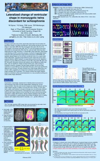
MZDZ. DSNR. DSDZ. DZNR. MZNR. MZDS. MZ. DS. DZ. NR. Left. Left. Right. Right. MZ. DS. DZ. 1. 3. 6. 10. Subjects and image data. Subjects : Image data provided by D. Weinberger, NIMH, Bethesda [2]: MZ : 10 healthy monozygotic twin pairs (N=2*10)

E N D
MZDZ DSNR DSDZ DZNR MZNR MZDS MZ DS DZ NR Left Left Right Right MZ DS DZ 1 3 6 10 Subjects and image data • Subjects: Image data provided by D. Weinberger, NIMH, Bethesda [2]: • MZ: 10 healthy monozygotic twin pairs (N=2*10) • DS: 10 MZ twin pairs discordant for schizophrenia (N=2*10) • DZ: 10 dizygotic twin pairs, all healthy controls (N=2*10) • NR: Selection of unrelated, healthy subject pairs with best possible match of age and gender (N=2*10) • Image data: Gradient-echo T1w (256x256x128, 240mm FOV, 1.5mm slice distance) Lateralized change of ventricular shape in monozygotic twins discordant for schizophrenia 2M Styner, 1,2G Gerig, 3DW Jones, 3DR Weinberger, 1JA Lieberman Dept. of 1Psychiatry and 2Computer Science University of North Carolina, Chapel Hill, NC 27614, USA 3 National Institute of Health, Bethesda, MD gerig@cs.unc.edu / http://www.cs.unc.edu/~gerig RESULTS Llateral ventricle shapes after volume normalization. Ventricles of 10 DS twin pairs (left) and 5 MZ and 5 DZ pairs (right) are shown side by side. Volume Analysis ABSTRACT Enlarged ventricular size and/or asymmetry have been found markers for psychiatric illness, including schizophrenia. We studied ventricular size and shape in volumetric MRI (N=2*30) of dizygotic twin pairs (DZ, N=2*10), monozygotic normal twin pairs (MZ, N=2*10), and monozygotic twin pairs discordant for schizophrenia (DS, N=2*10). Left and right ventricles were segmented from high resolution T1 SPGR MRI using automatic, atlas-based voxel labeling and 3D connectivity analysis. Surfaces of segmented lateral ventricles were parametrized using spherical harmonics (SPHARM) and spatially aligned based on the intrinsic coordinate frame of a coarse-scale shape description. Pairwise shape difference was measured as the mean squared distance (MSD) between corresponding surface points. Statistical analysis for group differences between normal MZ, discordant MZ (DS) and DZ using both, absolute volumes and relative volume difference within twin pairs, was not significant. Shape analysis within pairs, after scaling for individual volumes, revealed a strong shape similarity for left and right ventricles in normal MZ but also discordant DS pairs, which was significantly different from DZ pairs. DS pairs showed very small shape differences very similar to MZ, suggesting no change due to disease and strong pairwise shape similarity due to genetics. Our results indicate that shape analysis based on individual surface parameterization is a very sensitive technique to study subtle shape alterations. Relative ventricle volume differences between twin pairs (|T1-T2|/(T1+T2)) for the MZ, DS, DZ and NR groups. Mean difference tests were not significant between all the groups. Absolute volumes (top) and pairwise correlation of ventricle volumes (right). Global Shape Analysis Group differences of pairwise shape similarity (p-values) L R PROBLEM Ventricle shape differences between twin pairs for MZ, DS, DZ and NR groups. Shape difference metric: Mean square difference (MSD) between object surfaces. Quantitative morphologic assessment of individual brain structures inneuroimaging most often includes segmentation followed by volumemeasurements. Volumes and volume changes are intuitive features as they might explain atrophy or dilation of structures due to illness. On the other hand, subtle, well localized structural changes are not sufficiently reflected in global volume measurements. Development of new methods for three-dimensional shape analysis aims at tackling this issue and promises better sensitivity to subtle deformations. Shape analysis applied to twin studies offers the possibility to systematically study shape variability, both in healthy subjects and subjects discordant for disease, w.r.t. genetic difference. Locality: Average of local twin pair shape difference METHODS The T1w high resolution MR image data are processed by automatic brain tissue segmentation followed by shape parameterization. • MZ: Right ventricle between pairs more similar than left ventricle. • DS: Dissimilarity between pairs slightly larger than MZ but significantly smaller than DZ and NR. • DZ: Dissimilarity much larger than MZ and DS, but equivalent to NR. • Automatic, atlas-based 3D voxel segmentation technique. • Segmentation of lateral ventricles by manually guided 3D connectivity. Locality: Local group tests of pairwise differences Twin B Twin A • Parametrization of object surfaces using 3D Fourier harmonics (SPHARM. [1,3]). • Spatial alignment of structures by Procrustes fit of sets of homologous surface points. • Size normalization by individual volumes. • Pairwise shape difference: Mean square difference between surfaces (MSD). MZ • MZDS: No locality of subtle differences between pairwise shape dissimilarity. • DZNR: No locality of strong differences between pairwise shape dissimilarity. • MZDZ: Lateralization of locations of significant differences. Twin B CONCLUSIONS Twin A DZ • Analysis of volumes and volume differences: In general large volume variability and differences in all groups, but no significant differences between groups. • Pairwise shape differences after volume normalization: MZ DS < DZ = NR. • Using pairwise shape differences, MZ and DS are not significantly different. • Shape analysis adds information/sensitivity not provided by volume analysis. References: [1] A. Kelemen, G. Székely, and G. Gerig, „Three-dimensional Model-based Segmentation“, IEEE Transactions on Medical Imaging (IEEE TMI), 18(10):828-839, Oct 1999 [2] A. Bartley, D. Jones, and D. Weinberger, “Genetic variability of human brain size and cortical patterns”,Brain, vol. 120, pp. 257–269, 1997. [3] G. Gerig, M. Styner, D. Jones, D. Weinberger, and J. Lieberman, “Shape Analysis of brain ventricles using SPHARM”, in: Proc. Workshop on Math. Methods in Biomed. Image Analysis MMBIA 2001, IEEE Comp Soc, pp. 171-178, Dec. 2001 Hierarchical surface parameterization by SPHARM Parameterized object surfaces showing correspondences. February 2002