Download
1 / 48
480 likes | 499 Views
Discover the intricate anatomy of the ear, exploring its external, middle, and inner parts. Learn about its role in hearing and balance and how receptors and organs function within. Uncover the complexities of the auditory ossicles and the vestibular system. Dive into the details of the cochlea and the labyrinth's two divisions. Gain insights into static and dynamic equilibrium mechanisms in the inner ear.

E N D
I. Introduction to the EarA. Function: The Ear houses hearingand balance(equilibrium) 1. Receptors (Hair Cells) are mechanoreceptors • Sensory nerves are stimulated by bending hairs 2. Organs: Different ones inside ears for each sense Balance hearing
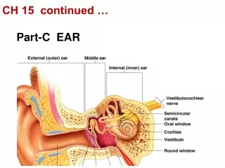
II. Anatomy: 3 areas of the ear • External (outer) ear • Middle ear (tympanic cavity) • Inner ear (bony labyrinth)
A. The External Ear = Auricle (Pinna) 1. Helix & Lobule: 2. External acoustic meatus (auditory canal) • Description: • Ceruminousglands • Tympanic membrane: Ear: Transfers sound wave to middle ear; Medial border of E.A.M.
B. The Middle Ear (Tympanic Cavity) – description = • 1. 3 Auditory Ossicles= malleus (hammer), incus (anvil), and stapes (stirrup) • Function: • 2. Pharyngotympanic(auditory) tube • Location: • Functions: • 3. Medial border = bony wall with 2 openings • a) round windows • b) oval window: Stapes fits on to it; transfers vibrations to inner ear Medial Border
Oval window (deep to stapes) Semicircular canals Entrance to mastoid antrum in the epitympanic recess Malleus (hammer) Vestibule Incu (anvil) Auditory ossicles Vestibular nerve Stapes (stirrup) Cochlear nerve Tympanic membrane Cochlea Round window Pharyngotympanic (auditory) tube (b) Middle and internal ear Figure 15.25b
4. Tensor tympani & Stapedius muscles: When contracted they reduce the vibrations of Auditory Ossicles; Function when sound is too intense Epitympanic recess Malleus Incus Superior Lateral Anterior View Pharyngotym- panic tube Tensor tympani muscle Tympanic membrane (medial view) Stapes Stapedius muscle Figure 15.26
C. Inner Ear or Labyrinth– hearing and balance 1. Introduction: Two Parts (Divisions) a) Bony Labyrinth = Hollowed out spaces surrounded by bone that contains the Membranous Labyrinth Parts: Cochlea, Vestibule &Semicircular canals b) Membranous Labyrinth = Made of tissue and containing sound and balance receptors. Shape resembles Bony Labyrinth, but slightly smaller so it can fit into it. Parts: • Cochlear Duct, Utricle &Saccule, Semicircular ducts SemicircularCanals w/ Semicircular Ducts Cochlea w/ Cochlear Duct Vestibule w/ Utricle &Saccule
2. VESTIBULE= egg-shaped bone cavity a) Saccule & Utricle of Membranous Labrinth • Functions: Static Equilibrium • Maculae = Sensory area containing hair cells • Monitors head position • Hair cells with stereo cilia are embedded in an Otolithic membrane • Otolithic membrane = jelly-like mass with calcium carbonate crystals (stones) b) Specific Functions: Static Equilibrium i) Utricle: hairs are vertical causing movement in the horizontal plain to bend hairs - Result: Horizontal Accelaration Macula of Utricle
ii) Saccule: hairs in macula are horizontal causing up and down movements to bend the hairs - Result: Vertical Acceleration c) Vestibular Branch of Vestibulocochlear Nerve Vestibular Branch Vestibulocochlear Nerve Cochlear Branch
Otoliths Kinocilium Otolithic membrane Stereocilia Hair bundle Macula of utricle Macula of saccule Hair cells Supporting cells Vestibular nerve fibers Figure 15.34
3. SEMICIRCULAR CANALS = cavities in bone • Function: Dynamic Equilibrium– Changes in velocity of rotational movement of head • Anterior, Posterior, Lateral • Semicircular Duct = Membranous Labrinth • Ampulla with Crista ampullaris
Superior vestibular ganglion Inferior vestibular ganglion Temporal bone Semicircular ducts in semicircular canals Facial nerve Vestibular nerve Anterior Posterior Lateral Cochlear nerve Cristae ampullares in the membranous ampullae Maculae Spiral organ (of Corti) Utricle in vestibule Cochlear duct in cochlea Saccule in vestibule Stapes in oval window Round window Figure 15.27
Crista ampullaris: contains hair cell receptors for equilibrium • Hair cells- in contact with cupula • Cupula = jelatinous strands that connect to the hair cells • Rotation causes fluid to go opposite direction moves hair cells • Result: Dynamic Equilibrium • Vestibular Branch, part of the Vestibulocochlear Nerve
Cupula Crista ampullaris Endolymph Hair bundle (kinocilium plus stereocilia) Hair cell Crista ampullaris Membranous labyrinth Supporting cell Fibers of vestibular nerve (a) Anatomy of a crista ampullaris in a semicircular canal Cupula (b) Scanning electron micrograph of a crista ampullaris (200x) Figure 15.36a–b
4. COCHLEA: chamber in bone that spirals around Modiolus a) Introduction • Modiolus– bony pillar • Three Membranous Labyrinth Chambers = Scalae: Scala Vestibuli, Scala Media, Scala Tympani • Cochlear Branch of Vestibulocochlear Nerve
Modiolus Cochlear nerve, division of the vestibulocochlear nerve (VIII) Spiral ganglion Osseous spiral lamina Vestibular membrane Cochlear duct (scala media) Helicotrema (a) Figure 15.28a
Vestibular membrane Osseous spiral lamina Tectorial membrane Spiral ganglion Scala vestibuli (contains perilymph) Cochlear duct (scala media; contains endolymph) Stria vascularis Spiral organ (of Corti) Scala tympani (contains perilymph) Basilar membrane (b) Figure 15.28b
b) ScalaVestibuli c) Media = COCHLEAR DUCT: houses Spiral Organ of Corti which has Hair cells • Vestibular Membrane: roof • Basilar Membrane: floor • Spiral Organ of Corti– in between above • Tectorial Membrane • Hair Cells with stereocilia embedded in tectorial membrane • Cells are moved when endolymph vibrates • Sensory (Afferent) Fibers of Cochlear Nerve Scala Vestibuli Cochlear Duct Scala Tympani
Organ of Corti d) Scala Tympani
III. Physiology– SoundA. HEARING—Properties of Sound 1. Sound is = A pressure disturbance produced by a vibrating object which pushes the air causing an area of high pressure first on one side of the object and then on the other side. As the air vibrates it also creates areas of low air pressure, alternating with the high air pressures. High Pressure & Low Pressure Wavelength Air Pressure
2. The alternating pressuresradiate out in all directions; much slower than light 3. Air molecules moves just a short distance 4. Molecules give off kinetic energy as they bump into the adjacent air molecules 5. The propagation: depends on the molecules of the medium (air); they must be elastic. 6. Sound Wave with a frequency and amplitude High Pressure & Low Pressure Wavelength Air Pressure
Area of high pressure (compressed molecules) Area of low pressure (rarefaction) Wavelength Air pressure Crest Trough Distance Amplitude A struck tuning fork alternately compresses and rarefies the air molecules around it, creating alternate zones of high and low pressure. (b) Sound waves radiate outward in all directions. Figure 15.29
6. Sound Wave with a frequency and amplitude … a) Frequency = Pitch • The number of waves that pass a given point in a given time • Wavelength: The distance between two consecutive crests • The higher the frequency, the shorter the wavelength the higher the pitch • Perception of different frequencies are in the range of 20–20,000 Hz; 1500-4000 best perceived
High frequency (short wavelength) = high pitch Low frequency (long wavelength) = low pitch Pressure Time (s) (a) Frequency is perceived as pitch. High amplitude = loud Low amplitude = soft Pressure Time (s) (b) Amplitude (size or intensity) is perceived as loudness. Figure 15.30
b) Amplitude = loudness • The height of the crests and is higher when there is more of a difference between the high pressure and low pressure • Loudness = Subjective interpretation of sound intensity • Normal range is 0–120 decibels (dB which are logarithmic scale: “2” is 10 times more intense than “1”) • Perception: The perceived loudness between two amplitudes is less than the actual difference • > 120 dB causes pain; can hear higher dB • > 90 dB causes damage depending on how often
B. Steps to HEARING: ) ) ) ) ) ) ) ) ) ) ) ) ) ) • Sound waves are amplified & transferred to the inner ear • Sound waves arrive at tympanic membrane. • Tympanic membrane vibrates the malleus (hammer) bone. • Vibrations amplified by middle ear bones.
Sound waves are transformed into hydrostatic pressure waves ) ) ) ) ) ) ) • Stapes (stirrup) bone vibrates oval window that creates fluid pressure waves inside vestibule. • Pressure waves travel down the cochlea. • Spiral organ of Corti inside cochleastimulated by pressure waves. Figure 8.12
7. hair receptors in organ of Corti bend from pressure waves and send a message (action potential) to the brain But how do we tell different notes from one another? ) ) ) ) ) ) ) ) ) ) ) ) Off to the brain for interpretation… Figure 8.15b
C. Auditory Processing1. Pitchis determined by where along the Cochlear Duct’s length that the membrane vibrates. High notes vibrate near entrance, low notes at end of cochlea ) ) ) Basal membrane ‘tuned’ to different frequencies;! Figure 8.16b–c
2. Loudness: is detected by increased numbers of action potentials (frequency of action potentials) that result when the hair cells experience larger deflections 3. Localization of sound: depends on relative intensity and relative timing of sound waves reaching the ear closest to the generation of the sound and the ear farthest away. The difference between what the ears hear indicated where the sound is coming from.
D. Homeostatic Imbalances of Hearing • Conduction deafness • Sensorineural deafness • Tinnitus: • Meniere’s syndrome:
IV. EQUILIBRIUM & ORIENTATION A. Vestibular Apparatus = receptors in Semicircular Canals and Vestibule about static and dynamic equilibrium • Vestibular nuclei in the Brainstem & Cerebellum • Receive input from vestibular apparatus, eyes, and somatic receptors • Function: brain can maintain balance & sense of movement and direction • Output from brain via reflexes to eyes and body muscles
Section of ampulla, filled with endolymph Fibers of vestibular nerve Cupula Flow of endolymph At rest, the cupula stands upright. During rotational acceleration, endolymph moves inside the semicircular canals in the direction opposite the rotation (it lags behind due to inertia). Endolymph flow bends the cupula and excites the hair cells. As rotational movement slows, endolymph keeps moving in the direction of the rotation, bending the cupula in the opposite direction from acceleration and inhibiting the hair cells. (c) Movement of the cupula during rotational acceleration and deceleration Figure 15.36c
B. Static Equilibrium – how we know up from down • Maculae—receptors in vestibule that sense head position • Pull of gravity causes otoliths (tiny stones) to bend hair cells • Utricle: Accelaration in horizontal plane & titling head to side • Saccule: Verticle accelaration 1 macula
C. Dynamic Equilibrium • Crista ampullaris—receptor organs in semicircular canals • Cupula (gelatinous cap) covers hair cells • Angular motion causes cupula to bend hairs = stimulate nerves
D. Equilibrium Pathway to the Brain • Pathways are complex and poorly traced • Impulses travel to the vestibular nuclei in the brain stem or the cerebellum, both of which receive other input • Three modes of input for balance and orientation • Vestibular receptors • Visual receptors • Somatic receptors
Otoliths Kinocilium Otolithic membrane Stereocilia Hair bundle Macula of utricle Macula of saccule Hair cells Supporting cells Vestibular nerve fibers Figure 15.34
Cupula Crista ampullaris Endolymph Hair bundle (kinocilium plus stereocilia) Hair cell Crista ampullaris Membranous labyrinth Supporting cell Fibers of vestibular nerve (a) Anatomy of a crista ampullaris in a semicircular canal Cupula (b) Scanning electron micrograph of a crista ampullaris (200x) Figure 15.36a–b
Participating… 5. Hearing and equilibrium are both sensed by _________________ that are stimulated by bending hairs. The outer and middle ear are involved in the sense of ___________ only, while the inner ear helps to sense both ____________ and _________________________. mechanoreceptors hearing hearing balance, i.e. equilibrium
Participating…#7 Sound waves cause the _________ membrane to vibrate which in turn causes the _________, _______, and _______ of the middle ear to transmit vibrations to the oval window of the inner ear. Pitch is then determined by where vibrations stimulate nerve cells inside the ___________. tympanic malleus (hammer) incus (anvil) stapes (stirrup) cochlea Figure 8.16a
Participating… 6. Static equilibrium is detected by tiny organs known as ___________ in the _________ of the inner ear. Angular motion, i.e. dynamic equilibrium is detected by tiny organs called __________ ____________ in the ______________ canals. maculae vestibule crista ampullaris semi-circular
c. Fluids: Perilymph & Endolymph Superior vestibular ganglion Inferior vestibular ganglion Temporal bone Semicircular ducts in semicircular canals Facial nerve Vestibular nerve Anterior Posterior Lateral Cochlear nerve Cristae ampullares in the membranous ampullae Maculae Spiral organ (of Corti) Utricle in vestibule Cochlear duct in cochlea Saccule in vestibule Stapes in oval window Round window Figure 15.27