Download
1 / 10
160 likes | 1.32k Views
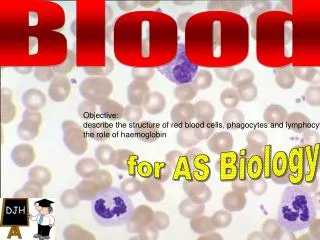
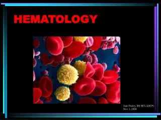
Granulocytes : Neutrophils/Eosinophils/Basophils. Classified according to cell morphology and cytoplasmic staining Neutrophils : stains with BOTH acid and basic dyes called ‘PMN’ for lobed nucleus; 50% of circ leukocytes

E N D
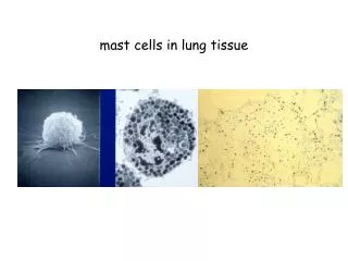
Granulocytes: Neutrophils/Eosinophils/Basophils • Classified according to cell morphology and cytoplasmic staining • Neutrophils: stains with BOTH acid and basic dyes • called ‘PMN’ for lobed nucleus; 50% of circ leukocytes • Eosinophils: stain with ACID dye (Eosin-red); bilobed nucleus; • 1-3% of leuko’s • Basophils: stain with BASIC dye (Methylene blue); <1% of • leuko’s
Neutrophils • Circulate in peripheral blood 7-10 hr before migrating into tissue; live only a few days • “front line of innate defense” • increased # (leukocytosis) used as an indicator of infection • extravasate in inflam rxn • attracted by chemotactic factors • active phagocytes; digestive enzyme held in 1° and 2° granules • Use both O2-dep and O2-indep digestive mech’s • Produce high levels of defensins
Eosinophils and Basophils Eosino’s • Like Neutrophils, function in phagocytosis • function vs. parasitic infections • contents of released granules damages parasitic membrane in ADCC • Non-phagocytic; function as “sirens” for inflam and allergy Baso’s
Dendritic cells (DC) • Resemble dendrites of nerve cells- hence the name • major role as APC TH • 4 types: Langerhans Interstitial DC’s Myeloid DC’s Lymphoid DC’s • all with hi levels of MHC II and B7 receptors • follicular DC’s localized to follicles and have receptors for Ab’s
Organs of the Immune system Primary lymphoid organs: location of lymphocyte maturation and immunocompetence *bone marrow *thymus gland Secondary lymphoid organs: locations where Ag contacts lymphocytes ranges from a) diffuse groupings of lympho’s and MØ (in lungs + lamina propria of int. wall) to b) lymphoid follicles – aggregates of lymphoid cells cradled by draining lymph vessels to c) lymph nodes and spleen – highly organized organs
Primary lymphoid organs: • Thymus Gland – flat, bilobed organ just above the heart • -surrounded by a capsule divided into lobules by strands of conn tissue called trabeculae • -each lobule has a peripheral portion Cortex • with immature T cells (thymocytes) + nurse cells • -each lobule has an inner portion Medulla • all is embedded in a stroma containing DC + MØ’s • *thymus generates T cells with large diversity of TCR’s; destroys those T cells which react with self Ag’s and those which cannot recog Ag/MHC
Thymic function declines with age; max size at puberty- atrophies thereafter
Primary Lymphoid Organs: 2) Bone marrow – not site of B dev in all species -site of both origin and development of B cells -stromal cells secrete cytokines req’d for B cell devlpt -those B cells which react to “self” are destroyed
Lymphatic system • BP serves to push plasma thru thin capillary walls • This “interstitial fluid” bathes cells of tissues • some returns to capillaries, some flows into lymphatic capillaries to lymphatic vessels to lymph nodes, etc to efferent lymph vessels to thoracic duct or right lymph duct where it (lymph) enters back into bloodstream and circulation • Foreign Ag gaining entry to lymph drainage is carried to local lymph nodes where it becomes trapped and presented to lymphocytes