Retinal Vascular Narrowing in Glaucoma: Intereye Comparison
Comparison of retinal arterial and venular caliber differences in fellow eyes with asymmetric glaucoma severity. Findings suggest retinal vascular caliber assessment as a potential indicator of glaucoma severity.

Retinal Vascular Narrowing in Glaucoma: Intereye Comparison
E N D
Presentation Transcript
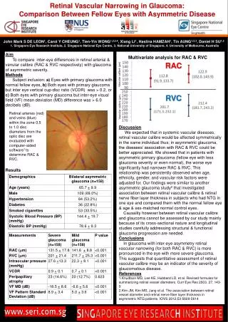
Retinal Vascular Narrowing in Glaucoma: Comparison Between Fellow Eyes with Asymmetric Disease John Mark S DE LEON1, Carol Y CHEUNG1, Tien-Yin WONG1,2,3,4, Xiang LI1, Haslina HAMZAH1, Tin AUNG1,2,3, Daniel H SU1,2 1. Singapore Eye Research Institute, 2. Singapore National Eye Centre, 3. National University of Singapore, 4. University of Melbourne, Australia Aim To compare inter-eye differences in retinal arterial & venular calibre (RAC & RVC respectively) with glaucoma of asymmetric severity. Methods Subject inclusion: a) Eyes with primary glaucoma with normal fellow eyes, b) Both eyes with primary glaucoma but inter eye vertical cup-disc ratio (VCDR) was > 0.2, or c) Both eyes with primary glaucoma but inter-eye visual field (VF) mean deviation (MD) difference was > 6.0 decibels (dB). Results Discussion We expected that in systemic vascular diseases, retinal vascular calibre would be affected symmetrically in the same individual thus; in asymmetric glaucoma, the diseases’ association with RAC & RVC could be better appreciated. We showed that in patients with asymmetric primary glaucoma (fellow eye with less glaucoma severity or even normal), the worse eye significantly had narrower RAC & RVC. This relationship was persistently observed when age, ethnicity, gender, and vascular risk factors were adjusted for. Our findings were similar to another asymmetric glaucoma study2 that investigated association between retinal vascular calibre & retinal nerve fiber layer thickness in subjects who had NTG in one eye and compared them with the normal fellow eye & age & sex-matched normal control eyes. Causality however between retinal vascular calibre and glaucoma cannot be assessed by our study mainly because of its cross-sectional nature and longitudinal studies carefully addressing structural & functional glaucoma progression are needed. Conclusions In glaucoma with inter-eye asymmetry retinal vascular narrowing (for both RAC & RVC) is more pronounced in the eye with more severe glaucoma. This suggests that quantitative assessment of retinal vascular calibre may be an indicator of the severity of glaucomatous disease. References Knudtson MD, Lee KE, Hubbard LD, et al. Revised formulas for summarizing retinal vessel diameters. Curr Eye Res 2003. 27: 143-9 Kim JM, Kim MS, Jang et al. The association between retinal vessel diameter and retinal nerve fiber layer thickness in asymmetric NTG patients. IOVS 2012;53:5609-5614 Multivariate analysis for RAC & RVC RAC Vascular calibre (µm) RVC Vascular calibre (µm) Retinal arteries (red) and veins (blue) within the zone 0.5 to 1.0 disc diameters from the optic disc are evaluated with computer-aided software1 to determine RAC & RVC.