Download
1 / 36
380 likes | 1.29k Views
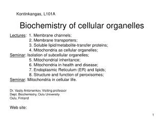
Cellular Basis of Biochemistry CELLS AND ORGANELLES (2h). Prof. KM Chan Department of Biochemistry Rm 513B, Basic Medical Science Building Tel: 3163-4420 Email: kingchan@cuhk.edu.hk. Table of Contents. Introduction The Cells The Organelles Summary Revision exercises.

E N D
Cellular Basis of BiochemistryCELLS AND ORGANELLES (2h) Prof. KM Chan Department of Biochemistry Rm 513B, Basic Medical Science Building Tel: 3163-4420 Email: kingchan@cuhk.edu.hk
Table of Contents • Introduction • The Cells • The Organelles • Summary • Revision exercises http://web.jjay.cuny.edu/~acarpi/NSC/13-cells.htm Cell: The most basic unit of life.
1. Introduction: 1.1 What is life? • Characteristics of life: life cycle; divide or die; grow or differentiate. • Pre-programmed message stored in genes: DNA makes RNAs make proteins (central dogma of life). • Viruses and phage rely on cells to replicate. Their genetic materials either DNA or RNA are wrapped within the protein coat. • Primitive cells might be developed from biomolecules/chemicals wrapped inside membrane. A cell membrane
1.2 How did the first cell emerge? • Life might be emerged in the ocean from lava rock during volcano outbreak when useful “chemicals” wrapped by membrane. • How was the first membrane formed? • Archaeon (Archae bacteria) found today have ether-lipids with branched chains to live in high temperature vent by volcano outlet.
1.3 Size Range • Small molecules (chemicals) < 1 nm (10-9 m) • Proteins and lipids- biomolecules < 10 nm • Ribosomes = 20 nm; viruses < 100 nm • Bacteria nm, 1 µm to 10 µm (10-6 m) • Mitochondrion, 2 µm; Nucleus, 7 µm • Plant and animal cells: 10-100 µm • Frog egg, 2 mm; salmon egg, 10 mm • Chicken egg, 4-5 cm • Length of muscle and nerve cells: 50-70 cm; multi-nucleated. Cells II: Cellular Organization http://www.emc.maricopa.edu/faculty/farabee/biobk/BioBookCELL2.html
2. The Cells2.1 The E. coli Cell and animal Cell Prokaryotic cells • They’re small and simple, it grows and divides fast. It can adapt to rapid environmental changes. • A single compartment with DNA and other biomolecules exist together in a well organized way. • Extra-chromosomal DNA exists in the form of plasmid. Eukaryotic cells • A hundred times larger, much more complex • Contain organelles to support different cellular functions from digestion to generation of energy and storage of nutrients. • Extra-chromosomal DNAs exist in mitochondria and chloroplast. • Differentiate to different cell-types.
Gram-negative cells are sensitive to pennicillin and lysozyme. Capsule contains lipo- polysaccharides (LPS) which are endotoxins to animals. Mesosome is infolding of membrane for specialized functions Capsule Cell wall Mesosome Membrane Gram-positive cell wall Gram-negative cell wall Nucleoid (DNA region ) capsule Periplasmic space Ribosomes peptidoglycan Lipo-protein outer membrane Flagella Peptidylglycan layers Amino acid layers membrane http://medic.med.uth.tmc.edu/slnk/00001467.gif
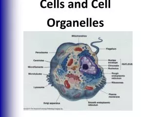
Model of Eukaryotic cell Schematic of typical animal cell, showing subcellular components. Organelles: (1) nucleolus (2) nucleus (3) ribosome (4) vesicle (5) rough endoplasmic reticulum (ER) (6) Golgi apparatus (7) Cytoskeleton (8) smooth ER (9) mitochondria (10) vacuole (11) cytoplasm (12) lysosome (13) centrioles http://en.wikipedia.org/wiki/Organelle
2.2 Cell wall and extra-cellular matrix • Bacterial cell wall is made of carbohydrates covering the outside of cell membrane. • Gram positive bacteria have simpler but a thick wall of peptidoglycan • Gram negative have less peptidoglycan, but more complex in structure with periplasmic space and lipopolysaccharides. • Plant cells also use cell wall to stabilize their structures. • Animal cells have extra-cellular (outside of the cell) matrix instead and they are more dynamics (being formed or disorganized quickly).
Compare (both are) Eukaryotic cells Contain organelles, for specific function Contain mitochondria Cytoskeleton (protein molecules) to hold up shape and structures Nucleolus in nucleus for rRNA synthesis Both contain rough endoplasmic reticulum 2.3 Animal cells and plant cells
Adapted from the Merck Manuals On-line Medical Library: http://www.merck.com/mmhe/sec01/ch001/ch001b.html#sec01-ch001-ch001b-6 Adapted from Molecular Expression (http://micro.magnet.fsu.edu/cells/plantcell.html) Animal cells develop to specific cell types in different organs (differentiations). Specific cell types are for specific biological functions (e.g. production and secretion of specific hormones or enzymes).
Contrast (the differences) Plants cells and fungal cells have cell wall which is made of carbohydrate Plant cells have mitochondria and chloroplast is only found in plants Vacuole and storage granules in plant cells for water control and nutrient storage Animal cells have lysosomes for digestion, microtubules, microfilaments, and centrioles for intracellular transport and cell division. Animal cells and plant cells
3. The organelles3.1 The Nucleus • Double membrane envelope that continues with endoplasmic reticulum. • Nuclear pores to communicate with outside of the nucleus. “Gating” is necessary to control factors for gene transcription and cell signaling to move around. • Luminal subunits in between the outer and inner membrane of the nucleus control the nuclear pores with a ring structure and nuclear cage. • Nucleolus is a structure inside the nucleus with chromatin actively transcribing ribosomal RNAs.
Nuclear translocator protein (vehicle) Architectures of nucleus Pore complex Outer membrane Inner membrane Laminal subunit Chromatin: DNA with histones and non-histone proteins Nuclear pores Ribosome: RNA translated to protein Inner membrane Outer Nuclear membrane Rough Endoplasmic Reticulum Nucleolus: ribosomal RNA genes being transcribed.
Genetic Information can be decoded by transcription Eukaryotes: genes have introns and exons Prokaryotes: polycistronic, co-translation exon mRNA Introns, intervening region (uncoding) AAAAA RNA splicing AAAAA Mature messenger RNA AAAAA Nucleus Cytoplasm Translation in rough endoplasmic reticulum or polysomes area Proteins made Polycistronic: one mRNA codes for several proteins/
3.2 Endoplasmic Reticulum (ER) • It spans the entire cell and attached with numerous ribosomes for protein translation (Rough ER, RER). • The proteins made in the RER are folded in proper conformation, may be glycosylated (post-translational modifications by adding sugars), and sorted to various parts of the cell. • Smooth ER (SER) produces phospholipids. • SER is a depot of calcium ions for cellular signalling.
Rough ER dedicated for protein production http://cellbio.utmb.edu/cellbio/ribosome.htm#Ribosome-Endoplasmic%20Reticulum From RER to Golgi Apparatus http://cellbio.utmb.edu/cellbio/golgi.htm
Protein synthesis and post-translational modification in RER; membrane proteins already fixed to the membrane surface. • Hydrophobic helical domains get through the membrane of RER. • Post-translational modification, e.g. glycosylation, sorting and delivery to proper places. • Orientations of these membrane proteins are kept. nucleus RER Golgi sugar Plasma membrane vesicle
3.3 The Golgi Apparatus • Proteins made in the RER are sorted and processed in the Golgi apparatus. • They enter the Golgi at the cis face from vesicles formed at the ends of the ER. • In the cisternae, proteins are labelled (secondary modification), sorted and delivered to the trans face of the Golgi apparatus. http://sun.menloschool.org/~birchler/cells/animals/golgi/structure.html
Tunicamycin (an antibiotic) • Proteins are glycosylated in RER before dispatch to the Golgi apparatus. • The antibiotic tunicamycin acts by mimicking the structure of UDP-N-acetylglucosamine, the substrate in the first enzymatic step in the glycosylation pathway. • It thus blocks protein post-translational modification and hence protein production is inhibited to kill eukaryotic cells.
Protein sorting (trafficking) • As vesicles formed in the Golgi and sent to proper locations for specific action in other organelles or stored as secretion vesicles. • Proteins in vesicles would accumulate and wait for signal to be exported out by exocytosis (either regulated or constitutively secreted out of the cells. • Some proteins are delivered to and kept on cell membrane. • Some special enzymes are sent to lysosomes for digestion.
Rough Endoplasmic Reticulum and Golgi Apparatus Lysosome Early endosome Rough Endoplasmic Reticulum Engulfed materials Regulated secretory pathways Vesicle Trans Golgi apparatus Constitutive secretory pathway Cis-Golgi apparatus Golgi Apparatus Proteins are further modified and sorted in the Golgi apparatus
Heterophagy Autophagy • The ER can engulf mitochondria or other internal organelles for recycling • Autophagosome formed after fusion of ER and mitochondria • Phagosome fused with primary lysosome • Secondary lysosome join to digest the mitochondria to form residual body • Biomolecules are recycled inside the cells • Endocytosis, engulfment of foreign compound or body • Phagocytic vacuole formed and become Phagosome • Primary lysosome get to the phagosome to form endosome • Secondary lysosome with acid hydrolases join the endosome for better digestion • Residual body remains; unwanted contents discharged by exocytosis.
3.4 The Lysosome • An intracellular digestive organ • Acidic condition to digest and remove unwanted materials or break down materials for re-use. • Digestive enzymes are deposited to primary lysosome from Golgi • The enzymes may be released outside of the cellular membrane or fused with autophagic vesicles to form phagocytic vacuoles (secondary lysosomes) or endosomes. http://micro.magnet.fsu.edu/cells/lysosomes/lysosomes.html
I- cell Disease & Lysosomal Hydrolases • Lysosomal hydrolases sent to the cis Golgi are covalently modified by adding Mannos-6-Phosphate (M6P). • M6P receptor recognizes the labelled hydrolases in the trans-Golgi and move them to the endosomes. • I-Cell disease is a genetic defect that produce the enzymes without M6P. Thus no enzymes sent to form endosomes for digestions. • Glycosaminoglycans and glycolipids accumulated (without proper digestions). Patients exhibit psychomotor retardation and skeletal deformities, they die early, usually before age 8.
Tay-Sachs Disease • A lysosome storage disease • Absence of specific lysosomal enzymes causes accumulation of unwanted cellular materials in the brain. • E.g. Mental retardation and blindness resulted from Ganglioside GM2 accumulation due to the lack of hexosaminidase A. • The lysosomes cannot function properly or even break to release not just digestive enzymes, but also “acid” to kill the cell.
Gout and Rheumatoid Arthritis • Gout is deposition of uric acid crystals of the joints often from over consumption of meat. • Other Rheumatoid factor complexes in the leucocytes (white cells) of the joints. • These rupturelysosome torelease enzymes that degrade the components of the synovial membrane in the joints, causing great pain and joint deformation.
3.5 The MITOCHONDRION • Two membranes: inner and outer. • The inner membrane folds to create the matrix space and forms inter-membrane space in between inner and outer membrane. • The matrix contains enzymes to oxidize metabolites • The inter-membrane has potential gradient generated by pumps at the inner membrane, energy released from the gradient produce ATP energy. This is the major function of mitochondria (energy production).
Outer membrane, with channel forming proteins but only permeable to 5 kDa molecules or less. Other enzymes here facilitate metabolism of lipid for use in matrix and transport of required protein in the mitochondrion. MAJOR FUNCTION OF MITOCHONDRION: OXIDATIVE PHOSPHORYLATION (oxidation of NADH to produce ATP) Matrix, internal space with enzymes for oxidation of pyruvate and fatty acids and for the citric acid cycle (Kreb’s cycle). Cristae in boxed region Inter-membrane space, for enzymes using ATP passing out of the matrix to phosphorylate other nucleotides. Inner membrane, folded into numerous cristae to increase total surface area for electron-transport chain (respiratory enzymes), ATP synthase and transporters for metabolites going through.
Endosymbiotic hypothesis of the origin of mitochondria • Mitochondria carry their own DNA and divide similarly to bacterial fission. • Some proteins to be used in mitochondria are imported from the cytoplasm translated from mRNAs made from nuclear genes. • The mitochondria and their “host” have a symbiotic relationship. • A billion years ago, aerobic bacterium was endocytosed (engulfed) by a primitive anaerobic eucaryotic cell, since then it was kept for the eukaryotic cells to produce energy. • Evidence are found from DNA sequences, codon usage, membrane contents and behaviors of mitochondria which are similar to those found in bacteria.
Mitochondrial Disease • Mitochondrial disease can arise from defective import of proteins (e.g. heat shock protein 70 needed for docking and folding of proteins in the mitochondria) or mutations in mitochondrial genes. • Tissues affected are those heavily dependent on ATP, e.g. nerve and muscle cells, causing mental retardation and muscular weakness. • Leber’s hereditary optic neuropathy and Kearns-Sayre Syndrome are caused by mutations of NADH-CoQ reductase gene; progressive blindness, abnormal heart beat and nerve degeneration resulted.
3.6 Peroxisomes • For oxidation of fatty acids and toxicants, it contains oxidative enzymes. • E.g. catalase in peroxisomes can decompose hydrogen peroxide into water. • Abundant in hepatocytes (liver cells), where oxidation of fatty acids (and other organic matters) takes place and produces hydrogen peroxide. • Life span is short and the numbers of peroxisome vary. • They replicate like mitochondria, and they are not budded from Golgi. The enzymes are sent and deposited into the peroxisomes.
Zellweger syndrome • Zellweger syndrome is caused by the mistakes of protein import into the peroxisomes. • Accumulation of long chain fatty acids and pristanic acids in plasma and tissues. • Causing severe impairment of many organs and death. http://academic.brooklyn.cuny.edu/biology/bio4fv/page/peroxi_s.htm
4. Summary: • Eukaryotic cells are highly organized with membranous organelle- isolated compartments for specific life processes and functions. • Organelles increase a cell’s available membrane space for numerous enzymatic reactions. • The nucleus contains DNA for gene transcription and production plus processing of RNAs. Transcriptional factors and signals go in and out through nuclear pores under a tight control. • Rough ER handles translation and post-translational modification; protein sorting and vesicle are formed in Golgi apparatus.
Summary (continued) • Genetic diseases are often found related to malfunction in protein sorting, processing or trafficking across organelles. • Lysosomes are for cellular digestions, peroxisomes are for fatty acid oxidation and detoxification. • Damages of lysosomes leading to release of digestive enzymes and many clinical symptoms. • Mitochondia are for respiration and ATP energy production; it has 2 membranes and its own DNA. They might be originated from endosymbiosis in primitive eukaryotic cells.
5. Revision Exercises • Compare and contrast prokaryotic and eukaryotic cells. • Compare and contrast plant and animal cells. • Explain the endo-symbiotic hypothesis of the origins of organelles in eukaryotes. • List all organelles and their functions. • What is nucleolus ? What is it for?