Download
1 / 22

E N D
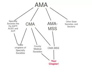
The assessment of the musculoskeletal system is challenging in pediatrics because of the variations in growth and development and the normal range of rotational changes in the extremities in infants and children. Increasing knowledge and comfort in the pediatric health care provider regarding the physical examination of the musculoskeletal system is key to accurate assessment, management, and refer-al of common orthopedic conditions.
Embryological Development • The rudimentary skeletal system forms as early as the fourth week of gestation when the devel- opment of the vertebrae begins and the upper extremities begin as buds on the fetus. As early as the seventh week of gestation, embryonic vascular growth progresses toward the center of the osteoblasts in the long bone, which forms cartilage. During the ninth week of ges- tation, ossification, or bone growth, begins in the ossification centers in the lower thoracic and upper lumbar vertebrae, and ossification continues in the femur.
The hand pads develop from the extremity buds by days 33 to 36, and the finger rays begin to form on days 41 to 43.1 It is during this period of gestation that polydactyly, the presence of extra digits, or syn- dactyly, the webbing or fusing of the digits, occurs along with other deformities of the extremities. The calcaneus, the largest tarsal bone of the foot, ossifies at the sixth month of fetal life. The position of the fetus in utero has the greatest impact on the skeletal system and the most common variants present at birth. • Muscle structures, including the tendons, ligaments, cartilage, and joints, originate from the embryonic mesoderm. Muscle fibers are developed by the fourth or fifth month of ges- tation. They increase in size along with changes in muscle and fat proportions throughout childhood and adolescence.
Developmental Variations • At birth, the epiphyses of the long bone are composed of hyaline cartilage. Shortly after birth, the secondary ossification centers begin to replace the cartilage along the epiphyseal plate. During early periods of rapid growth, os- sification occurs in secondary sites throughout the body: the ends of the long bones, the verte- brae, the flat bones in the clavicle, and the skull. The replacement of cartilage by bone is known as endochondral ossification. The epiphyseal plate ossifies, becoming the diaphysis, the shaft of the long bone; and cartilage at the metaphyseal plate replaces bone cells
Bone growth continues in infants and chil- dren along the epiphysis as new bone is added to the outer surface of existing bone. Growth along the epiphyseal plate continues until the cells in the growth plate stop dividing in puberty, and closure of the growth plate occurs in young adults when the metaphysis and the epiphysis fuse. • Several factors influence the health of the bones and growth of cells at the epiphyseal plate. Trauma during childhood can cause sep- aration of the epiphysis and the blood vessels to rupture, resulting in cessation of bone growth and a shortened extremity.1 Nutritional factors such as adequate protein in the diet, the amount of calcium intake, and adequate intake of vitamin D, particularly in breastfed infants, impact bone growth.
Vitamin D absorbs dietary calcium and phosphorus from the intes- tines and promotes bone health by maintain- ing normal levels of these minerals in the blood. Adequate levels of vitamin D in child- hood may also have a role in improving muscle and immune function. Parents are often ad- vised to keep infants and children out of the sun due to long-term risks of skin cancer, therefore reducing vitamin D synthesis from the skin. Maintaining adequate levels of vita- min D in infants and children is important to maintaining bone strength. Rickets is a defi- ciency of vitamin D in which the growth plate is impaired by calcification of newly formed bone on the metaphyseal plate. Alterations in thyroid or growth hormones can also impact normal growth.1 Recent evidence indicates environmental and chemical hazards may be a contributing factor to malformations in the musculoskeletal system.
Upper Extremities and Torso • The clavicle lies in a horizontal plane above the first rib and rotates between the sternum and the superior surface of the scapula. The scapula, a large, flat triangular bone, forms the posterior portion of the upper extremity and the superior, lateral angle articulates with the capsule or rounded head of the humerus, the large bone in the upper arm. The distal end of the humerusforms the condyle and is divided into the medial epicondyle and the lateral epicondyle. The proximal end of the ulna attaches at the articulating surface of the humerus and forms one of two long bones in the forearm. The olecranon is the outer, rounded distal surface of the ulna and forms the major portion of the elbow. The distal end of the ulna is small and articulates with the wrist bones. The radius is along the lateral side or thumb side of the fore- arm, and the distal end of the radius forms a large portion of the wrist bones.
The hand consists of eight carpal and five metacarpal bones. Many of the carpal bones are cartilaginous at birth. A radiograph of a child’s hand at 21⁄2 years of age illustrates ossification of only the capitate and hamate bones in the hand. Development and ossification of the hand continues until 11 years of age when all of the carpal bones are ossified except for the small pisiform bone, which develops by 12 years of age. Abnormalities of the phalanges, or fingers, such as syndactyly, webbing, or fusion between adjacent digits of the hands or feet, may be indicative of profound develop- mental delay. Polydactyly, the presence of supernumerary digits, is a more common variant and is not usually associated with other congenital anomalies.
Spine • The infant has a C-shaped spinal curve at birth in comparison to the double S curvature • that is present in late adolescence. Thoracic and pelvic curves are present at birth, and the secondary cervical curve is present by 3 to 4 months of age when the infant begins to hold up his or her head. The lumbar curva- ture begins to form as the infant bears weight and begins to walk. The young child often has an exaggerated thoracic-lumbar curvature and a protuberant abdomen until 3 years of age, when gait and balance become more normal . The sacrum is composed of five separate bones at birth and by 18 to 20 years of age is fused into one large bone.
The coccyx consists of three or four small bones that begin to ossify between the first and fourth years of life and fuses into one bone by 25 years of age • Pelvis • The hip bone contains three distinct parts in childhood, which are later fused in adulthood. The ilium is the superior, broad, flat surface of the hip bone. The ischium is the strongest • portion of the hip bone; it contains a portion of the acetabulum, which forms the attach- ment at the hip bone for the femur and the opening or foramen. The pubis contains the medial portion of the acetabulum and joins medially to form the complete pelvic girdle.
Lower Extremities and Feet • The femur is the longest and strongest bone in the body. The head of the femur becomes ossified during the first year of life, but the shaft of the femur does not become completely ossified until 14 years of age. The long bones of the extremities continue to grow at the site of the epiphyseal plate throughout childhood and adolescence; peak bone mass is achieved by young adulthood. In the lower leg, the larger bone is the tibia, the proximal end articulates with the femur and the distal end forms the medial malleolus of the ankle. The fibula is the smaller bone in the lower leg and lateral to the tibia. The distal end of the fibula forms the lateral malleolus of the ankle.
The patella is the small triangular bone that forms the kneecap and lies over the junction of the femur and the tibia. The health of the patella during growth and development depends not only on strong bone growth but also on the strength of the ligaments and tendons supporting the patella. • The seven tarsal bones and five metatarsal bones of the foot undergo dramatic ossification during the first year of life. The ossification of the bones in the foot follows the normal development of the gross motor milestones. The talus is ossified by the seventh month of life to form the ankle, and the remaining metatarsal bones continue to form during the latter half of the first year of life. The phalanges continue the ossification process through adolescence.
The limbs go through a continuous pro- cess of rotational change until about 8 years of age. The torsional development is a pro- cess that begins in infancy and progresses from sitting position to pulling up, to crawl- ing, then standing, cruising, and finally walking with an exaggerated gait and a wide base of support. The laxity in the ligaments and the torsional forces in the intrauterine environment as well as sitting and sleeping patterns in the early years are responsible for the normal variations in the lower extremities during childhood. Torsional variations • and abnormalities are hereditarily linked and often show a strong family tendency.
Monitoring Growth • During growth, bone has the remarkable capacity to remodel itself. Bone is deposited in areas subjected to stress and reabsorbed in areas where there is little stress. This physio- logical process explains the ability of children to remodel residual deformity after fractures and explains the biological plasticity in grow- ing bones. The rate of bone growth is greatest in the lower extremities before the onset of puberty; the distal extremities reach adult size in early puberty, which is reflected in shoe size in preadolescents. The trunk and proximal extremities exhibit the dominant growth dur- ing puberty and into young adulthood. Bone growth is completed at age 20 years, and peak bone mass is achieved by age 35 years. • Monitoring the velocity of growth is the key focus of routine well-child visits and when following children with chronic health conditions. For the child with slow growth or delayed onset of puberty, obtaining past growth charts and evaluating the progression of the growth curve along with familial patterns of growth and pubertal development is critical to assessing an abnormal growth pattern.
Muscle Development • The rate of muscle growth and development in- creases rapidly beginning at age 2 years. Early muscle growth is balanced by a normal decrease in adipose tissue in early childhood.1 The growth and maturation of the muscles continues with the ossification of the skeletal system. • Muscle tone is the state of normal tension of the muscles, and term infants normally have strong muscle tone at birth. Abnormalities in muscle tone in an infant may be noted shortly after birth when an infant is floppy. Less than normal muscle tone is described as hypotonia; rigidity or increased muscle tone is known as hypertonia; and spasticity is resistance to flexion and extension and range of motion of the muscles. Normal muscle function requires the normal function of the lower motor neurons (LMNs) in the spinal cord and the normal reflexivity of the muscle fibers. Normal muscle tone requires the normal function of the spinal cord stretch reflex and the balance of the upper motor neurons (UMNs) and LMN function. Lesions on the UMNs result in increased tone and LMN lesions result in decreased tone. • The greatest increase in muscle size occurs during puberty when boys’ muscle mass and muscle cell size begins to exceed that in girls. In early and middle childhood, girls’ muscle size increases at a greater rate than in boys up until puberty.
Examination of skeletal system • Check: - the shape of the head (it must be rounded) • skull deformations or abnormal head shape (and availability (occurrence, presence) head molding orcephalhaematomas) • state of fontanelle and sutures, • ratio of brains and face • state of orbits, nasal septum • number of teeth, bite • During a head first birth, pressure on the head caused by the tight birth canal may "mold" the head into an oblong rather than round shape. • This is a common occurrence that usually disappears after a few days. • Newborn head molding is a common occurrence that usually disappears after a few days. • The main sutures. (sagittal, coronal and occipital) must be close by the age of 3-4 m. • Early or late closure of any one or more of these sutures will result in an abnormal head shape, as shown below • Abnormal head shape • An abnormally shaped head is usually recognized at birth. • There are three causes of abnormal head shape in infants. • The largest group of infants with an abnormal head shape is those who have positional deformities which develop during pregnancy or while sleeping. • The next most common group is those infants who present with early closure of the cranial sutures (craniosynostosis). • The smallest group are infants with craniofacial syndromes, such as Apert's, Crouzon's, and Pfeiffer's.
Micrognathia, or "small jaw", can often be best appreciated when looking at the profile of the infant. • While many infants may appear to have a slightly recessed chin, the appearance above is clearly abnormal. • Underlying genetic conditions should be considered in cases such as this; • there are quite a few chromosomal conditions associated with this finding. • Down's syndrome is clearly the most common and recognizable genetic syndrome known, although at times the physical features may be difficult to identify in a newborn. • A typical facial features of Down's syndrome are: upslanting palpebral fissures, a suggestion of epicanthal folds, and a flat nasal bridge. • Cleft palate is not always this obvious. • When examining the palate, the examiner should palpate both the hard and soft palate to check for submucosal and partial clefts, which are more easily missed. • While examining chest, it’s necessary to check its shape (columnar, cone-shaped,barrel-like). • It’s necessary to pay attention on chest deformations: keeled chest, funnel chest; Harrison sulcus, heart hump. • Scoliosis is lateral curvature of the spine, which indirect signs are: • chest asymmetry – irregular disposition of shoulder – blades and shoulders, • different level of papillas, • smoothing out of waist triangles. • The forward bend test is a test used most often in schools and doctor's offices to screen for scoliosis. • During the test, the child bends forward with the feet together and knees straight while dangling the arms. • Any imbalances in the rib cage or other deformities along the back could be a sign of scoliosis. • It’s necessary to examine child’s limbs in standing and lying position: check their shape, ratio of limbs length, deformation (shortening, clubfoot), quantity of fingers, congenital dislocation of hip, dysplasia of bone system. • You must check the shape of joints, their deformations, skin colour, limitation of joint mobility, hypermobility, which is the sign of some hereditary dysplasias of connective tissue.
Palpation • With the help of palpation you must define integrity , density and painfulness of skull bones, the state of fontanelles and sutures. On pressure over the occipital bone you can define its pathologic osteomalacia – craniotabes. This is a symptom of rickets. • Early closing of big (anterior) fontanelle can occur in case of hypervitaminosis D or microcephaly. • Late closing of big fontanelle happens in case of rickets or hydrocephaly. • Increasing of tension and bulging fontanelle indicate raised intracranial pressure. When tension is increased very much, the pulsation stops. • Sunken fontanelle indicates dehydration. • While palpating bones and joints you must observe the child’s behaviour because during some diseases palpation can be painful. • It’s necessary to define temperature, sensitivity, thickness and mobility of the skin over the joints, its thickenings, edemas. • It’s important to check the presence of effusion into joint space by the fluctuation technique and floating patella symptom. Joint mobility is checked in all directions of (axes) joint movement.
Examination of muscular system: • Complaints, case history facts according to the child and his parents, inspection, palpation, If necessary instrumental research • Complaints: • muscular weakness (myasthenia), limitation of movements, muscular pains (myalgia). • Inspection • detect symmetry of muscles, degree of muscle development, presence of atrophy of muscular tissue, palsies and paresis, gained muscular deformities, congenital anomalies and assessment of muscular tonus • There are 3 degrees of muscle development: • 1.Weak muscle development – dropping abdomen, asymmetrical shoulder blades, and winged scapulas angle of scapula protrude slightly from body. Body and limb muscles at rest and on exercise are not contoured. • 2. Moderate development – abdomen is convex or flat, body muscles at rest are marked moderately, limb muscles are well-marked. While straining the muscles change shape. • 3. Good development – flat abdomen, scapulas are symmetrical, body and limb muscles at rest are well-marked, while straining there is a distinct shape of contracted muscles. • Muscle tone is a definite rigidity (tense) of skeletal muscles at rest.
Pathologic disorder of muscle tone is observed in children with disorders of nervous system and when there are dystrophic-degenerative changes in muscles because of metabolic disorders, toxic and other pathologic disturbances • The assessment of tone can be made both from observing the posture and activity of the infant when undisturbed and also by handling the baby. • Muscle tone indicates the ability of a muscle to respond to a stretch • Muscle tone is the amount of tension or resistance to movement in a muscle. • It is not the same as muscle weakness, which is a reduction in the strength of a muscle, but it can co-exist with muscle weakness. • Muscle strength in infants is tested by the degree of strain, necessary for opposition of any movement of the child. For example, try to take away a toy from the child. • Muscle strength of older children is tested by: • 1. The strength of handshake; • 2. Ability to resist the doctor when he flexes and extends limbs.