Download
1 / 15
200 likes | 619 Views
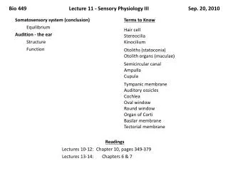
Bio 449 Lecture 11 - Sensory Physiology III Sep. 20, 2010. Readings Lectures 10-12: Chapter 10, pages 349- 379 Lectures 13- 14 : Chapters 6 & 7. Somatosensory system (conclusion) Equilibrium Audition - the ear Structure Function. Terms to Know Hair cell Stereocilia Kinocilium

E N D
Bio 449 Lecture 11 - Sensory Physiology III Sep. 20, 2010 Readings Lectures 10-12: Chapter 10, pages 349-379 Lectures 13-14: Chapters 6 & 7 • Somatosensory system (conclusion) • Equilibrium • Audition - the ear • Structure • Function Terms to Know Hair cellStereociliaKinocilium Otoliths (statoconia) Otolith organs (maculae) Semicircular canalAmpullaCupula Tympanic membraneAuditory ossiclesCochleaOval windowRound windowOrgan of CortiBasilar membraneTectorial membrane
Hair Cells (a) At rest (b) Excitation (c) Inhibition Tip link Channels closed.Less cation entryhyperpolarizes cell. Somechannelsopen More channelsopen.Cation entrydepolarizescell. Stereocilium Hair cell Primarysensoryneuron Action potentials Action potentials increase No action potentials mV Action potentials inprimary sensory neuron Time 0 mV –30 Release Release Membrane potentialof hair cell Excitation opension channels Inhibition closesion channels Fig. 10-22
Anatomy of the Ear EXTERNAL EAR MIDDLE EAR INNER EAR Semicircularcanals Nerves Cochlea Vestibularappartus Fig. 10-18
Vestibular Apparatus SEMICIRCULAR CANALS Superior Cristae withinampulla Horizontal Posterior Cochlea Utricle OTOLITH ORGANS Saccule Maculae Fig. 10-25
Kinocilium Stereocilia Otolith organs Otoliths Gelatinous layer Hair cells Supporting cells Similar toFig. 10-25 Sensory nerve fibers
Vestibular Apparatus SEMICIRCULAR CANALS Superior Cristae withinampulla Horizontal Posterior Cochlea Utricle OTOLITH ORGANS Saccule Maculae Fig. 10-25
Orientation of Semicircular Canals Vestibularapparatus Superior canal(nod for “yes”) Posterior canal(head tilt) Left right Horizontal canal(shake headfor “no”)
Endolymph Movement Cupula Bone Endolymph Hair cells Bone Direction of rotation of the head
Anatomy of the Ear EXTERNAL EAR MIDDLE EAR INNER EAR The pinnadirects soundwaves intothe ear Ovalwindow Malleus Incus Nerves Stapes Cochlea Earcanal Tympanicmembrane Roundwindow Topharynx Eustachiantube Fig. 10-18
Anatomy of the Inner Ear Ovalwindow Vestibularduct Cochlearduct Organ ofCorti Saccule Cochlea Uncoiled Helicotrema Roundwindow Tympanicduct Basilarmembrane
Anatomy of the Inner Ear Bony cochlear wall Vestibular duct Cochlear duct Tectorial membrane Organ of Corti Cochlear nerve transmitsaction potentials fromthe hair cells to theauditory cortex. Basilarmembrane Tympanicduct
Transmission of Sound Cochlear nerve Incus Ovalwindow Malleus Stapes Ear canal 5 Vestibular duct(perilymph) 3 Cochlear duct(endolymph) 2 6 4 1 Tympanic duct(perilymph) Tympanicmembrane Roundwindow
Transmission of Sound The movement of the tectorialmembrane moves the cilia onthe hair cells. Fluid wave Cochlearduct Tectorialmembrane Haircell Tympanicduct Nerve fibers ofcochlear nerve Basilar membrane