Download
1 / 77
1.01k likes | 2.31k Views
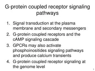
G-protein coupled receptor signaling pathways. Signal transduction at the plasma membrane and secondary messengers G-protein coupled receptors and the cAMP signaling cascade GPCRs may also activate phosphoinositides signaling pathways and produce calcium transients

E N D
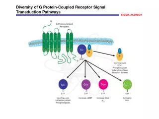
G-protein coupled receptor signaling pathways • Signal transduction at the plasma membrane and secondary messengers • G-protein coupled receptors and the cAMP signaling cascade • GPCRs may also activate phosphoinositides signaling pathways and produce calcium transients • G-protein coupled receptor signaling at the genome level
1. Signal transduction at the plasma membrane and secondary messengers • Cell-cell communication in a pluricellular organism • Signal transduction at the plasma membrane and secondary messenger production • The four classes of cell surface receptors
Chemical signaling from cell to cell Hormones Neurotransmitters Growth factors Extracellular matrix Cell adhesion Guidance molecules Recognition molecules
Diffusion-mediated cell to cell communication autocrine signaling paracrine signaling gap junctions Neurotransmitters Eicosanoides (prostaglandines, leukotrienes), Ca2+, small molecules Growth factors Quorum sensing Development D (µm2.s-1) r@1msec (µm) r@1sec (µm) Water 3000 1.7 55 Na+ 1000 1 32 acetylcholine 200 0.5 14 insulin 150 0.4 12 Diffusion kinetics : r2(t) = Dt
Cell to cell communication using active transport Endocrine system : secreting cells + specific receptors Neuronal system : impulse transmission + transmitter release Soluble hormones, Growth factors neurotransmitters 1-10 min 1-100 msec
Receptors, primary and secondary messengers Primary signal : hydrophilic signaling molecule Primary signal : hydrophobic signaling molecule Second messenger production Soluble hormones, growth and differentiationfactors, neurotransmitters, neurotrophins, extracellular matrix components, cytokines, chemokines, pathogen associated molecular patterns (PAMP) Steroid hormones, vitamin D, retinoic acid, nitric oxide (NO), aromatic hydrocarbone molecules, pathogen associated molecular patterns (PAMP)
1 The four classes of cell-surface receptors 907 G-Protein Coupled Receptors 2 3 109 Cytokine receptors About 400 sequences code for ion channel subunits 4 6 Guanylate Cyclase Receptors ? Receptors with phosphatase activity 58 Tyrosine Kinase Receptors Phosphatase activity
Intracellular messengers • Ion entry through ion channel activation : • Na+, K+, Cl- : induce plasma membrane voltage changes • Ca2+ : bind specific intracellular targets, activate PKC, CaM-K • Synthesis of small molecules : • cAMP, cGMP nucleotides : bind PKA or ion-channels • PIP2, AA phospholipids and derivatives : recruit proteins at the plasma membrane, activate PKB, PKC • Kinase or phosphatase activity : • Phosphorylation or dephosphorylation of specific aminoacids changes the conformation of proteins or allows their binding to other partners phosphorylation dephosphorylation
H NH C CO H CH3 C OH H H H NH C CO NH C CO HN C CO H H H H C C CH2 O OH O OH P O- OH Amino acid phosphorylation Threonine Thr, T Phosphorylated serine Ser-P, S-P Serine Ser, S + ATP + ADP + H+ Tyrosine Tyr, Y • Serine (S), Threonine (T) and Tyrosine (Y) are the main amino acids that can be phosphorylated. Kinase specificity is determined by the surrounding sequence (consensus sequence) • Phosphorylation modifies the H bonding network around the modified amino acid. • Phosphorylation adds a negative charge to the modified amino acid. • Phosphorylation can be mimicked by mutation to a negatively charged amino acid, such as aspartate (D) or glutamate (E). • Phosphorylation can be prevented by mutation to an amino acid lacking the hydroxyl group : S/T A, Y F • In bacteria, Histidine (H) can also be phosphorylated
2. G-protein coupled receptors and the cAMP signaling cascade • cAMP production : heterotrimeric G-protein activation and stimulation of adenylate cyclase activity • Example of the adrenergic receptor family • G-protein activation • Protein Kinase A (PKA) activation • Example of immediate responses : ion channel activation of olfactory neurons • Example of fast responses : glycogen degradation • Example of delayed signal transduction to the nucleus : the cAMP response element and CREB • GPCR receptor desensitization by specialized kinases and arrestins • Receptor downregulation at the plasma membrane
Amplification Regulators of G-proteins Stimulation (RGS) - b-Adrenergic Receptor Kinase (BARK) + Arrestin A specific receptor kinase phosphorylates a cytoplasmic loop of the receptor, allowing binding of an arrestin protein that prevents G protein activation
Cerebral brain cortex Somatic nervous system Autonomic nervous system Hypothalamus Heart rate Blood pressure (vascular tension) Hunger, thirst Circadian cycles Body temperature Sex organ function Breast milk production Water and osmolarity regulation Vasopressin CRH Pituitary gland (hypophyse) ACTH ‘sustained stress response’ Adrenal gland Corticosteroids (cortisol, DHEA dehydroepiandrosterone) Catecholamines (epinephrin) by chromaffin cells cortisol epinephrine ‘fight or flight response’
The structure of b-adrenergic receptors • Adrenergic receptors bind catecholamines, especially epinephrine (adrenaline) and norepinephrine (noradrenaline). There are 6 a- and 3 b-subtypes, characterized by their relative sensitivity to different agonists and antagonists. • These receptors have 7 transmembrane spanning domains. The binding of an agonist from outside the cell induces a conformational change that allows G-protein activation inside the cell. • Antagonists bind to the adrenergic receptors but do not trigger any response and furthermore prevent agonist-induced responses. A b-2 agonist :adrenaline outside Molecular structure of the b-2 adrenergic receptor and bound epinephrine (PDB 2rh1) A b-2 agonist :noradrenaline plasma membrane A b-2 antagonist : propanolol inside
Catecholamine effects • Effects of epinephrin • Acceleration of heart and breath rates • Dilatation of blood vessels in muscles and heart • Increase of glucose and fatty acid content in blood • Acceleration of instantaneous reflexes • Dilation of pupil • Inhibition of smooth muscles in the digestive track • Constriction of blood vessels in viscera • Transiently suppress the immune system • Effects of norepinephrin • Dilatation of blood vessels in muscles and heart • Increases attention (norepinephrin synthesized in the brain)
outside N Human beta-2 adrenergic receptor sequence Swiss-Prot P07550 extracellular 10 20 30 40 50 60 MGQPGNGSAF LLAPNGSHAP DHDVTQQRDE VWVVGMGIVMSLIVLAIVFGNVLVITAIAK 70 80 90 100 110 120 FERLQTVTNY FITSLACADLVMGLAVVPFGAAHILMKMWT FGNFWCEFWTSIDVLCVTAS 130 140 150 160 170 180 IETLCVIAVD RYFAITSPFK YQSLLTKNKA RVIILMVWIVSGLTSFLPIQMHWYRATHQE 190 200 210 220 230 240 AINCYANETC CDFFTNQAYAIASSIVSFYV PLVIMVFVYS RVFQEAKRQL QKIDKSEGRF 250 260 270 280 290 300 HVQNLSQVEQ DGRTGHGLRR SSKFCLKEHK ALKTLGIIMGTFTLCWLPFF IVNIVHVIQD 310 320 330 340 350 360 NLIRKEVYILLNWIGYVNSGFNPLIYCRSP DFRIAFQELL CLRRSSLKAY GNGYSSNGNT 370 380390 400 410 GEQSGYHVEQ EKENKLLCED LPGTEDFVGH QGTVPSDNID SPGRNCSTND SLL inside transmembrane domain agonist binding cytoplasmic
http://www.ch.embnet.org/software/TMPRED_form.html H1 H2 H3 H4 H5 H6 H7 Hydrophobicity (transfert energy) Amino acid position
The two conformations of heterotrimeric G proteins G G subunit : GDP/GTP binding G subunit : receptor binding G subunit : membrane binding GTP GDP
Demonstration of functional domains in G protein – coupled receptors by experiments with chimeric proteins containing portions of the β2- and α2-adrenergic receptors. Xenopus oocytes microinjected with mRNA encoding the wild-type receptors or chimeric α-β receptors expressed the corresponding receptor protein on cell surfaces. Although Xenopus oocytes do not express adrenergic receptors, they do express G proteins, which can couple to the foreign receptors. Binding assays were conducted using agonists known to bind selectively to α or β receptors to determine the ligand-binding specificity of the chimeric receptors. The effects of the agonists on adenylyl cyclase activity were taken as a measure of whether the receptor protein bound to the stimulatory (Gs) or inhibitory (Gi) type of oocyte G protein. A comparison of chimeric receptor 1, which interacts with Gs, and chimeric receptor 3, which interacts with Gi, shows that the G protein specificity is determined primarily by the source of the cytosol-facing loop between α helices 5 and 6. A comparison of chimeras 1 and 2 indicates that α helix 7 plays a role in determining the ligand-binding specificity. [See B. Kobilka et al., 1988, Science240:1310; W. A. Catterall, 1989, Science243:236.] Lodish Molecular Cell Biology
outside N Human beta-2 adrenergic receptor sequence extracellular 10 20 30 40 50 60 MGQPGNGSAF LLAPNGSHAP DHDVTQQRDE VWVVGMGIVMSLIVLAIVFGNVLVITAIAK 70 80 90 100 110 120 FERLQTVTNY FITSLACADLVMGLAVVPFGAAHILMKMWT FGNFWCEFWTSIDVLCVTAS 130 140 150 160 170 180 IETLCVIAVD RYFAITSPFK YQSLLTKNKA RVIILMVWIVSGLTSFLPIQMHWYRATHQE 190 200 210 220 230 240 AINCYANETC CDFFTNQAYAIASSIVSFYV PLVIMVFVYS RVFQEAKRQL QKIDKSEGRF 250 260 270 280 290 300 HVQNLSQVEQ DGRTGHGLRR SSKFCLKEHK ALKTLGIIMGTFTLCWLPFF IVNIVHVIQD 310 320 330 340 350 360 NLIRKEVYILLNWIGYVNSGFNPLIYCRSP DFRIAFQELL CLRRSSLKAY GNGYSSNGNT 370 380390 400 410 GEQSGYHVEQ EKENKLLCED LPGTEDFVGH QGTVPSDNID SPGRNCSTND SLL inside transmembrane domain agonist binding G-protein activation cytoplasmic
[RL] [L] [RL] Bmax - [RL] [L] K Reciprocal interactions between agonist and G-protein on the receptor • In the absence of guanine nucleotide, the agonist affinity is apparently higher : the ‘high affinity’ state correspond to Ragonist-Gempty complexes. Absence of nucleotide prevents agonist dissociation from the receptor. This is not a true equilibrium ! • In the presence of agonist, the ‘apparent’ GTP affinity for G-protein is increased and GDP affinity is decreased. This corresponds to a steady-state situation (and not to a true equilibrium) as Ga-GTP does not release its bound G(T/D)P before the next activation cycle. Scatchard plot : R + L RL affinity K K = and [R] + [RL] = Bmax K + [RL] = Bmax = [R].[L] [RL] B/F B
Competition binding isotherms of isoproterenol ([125I]ICYP = iodocyanopindolol = I-labeled isoproterenol) on membranes derived from HEK-293 cells expressing the b1-AR. The competition isotherms were determined in either the absence (-GTP) or presence of 100 µM Gpp(NH)p (+GTP). Zeitoun et al. (2006) Mutagenesis within Helix 6 of the Human 1-Adrenergic Receptor Identifies Lysine324 as a Residue Involved in Imparting the High-Affinity Binding State of Agonists Mol Pharmacol 70:838-850 log( ) Scatchard plot of cannabinoid activation of [35S]GTPγS binding in rat cerebellar membranes, showing basal (open symbols) and activated (closed symbols) [35S]GTPγS binding, as determined with 3 µM WIN 55212-2 (a cannabinoid agonist). Inset shows net agonist-stimulated [35S]GTPγS binding, with a high affinity of 2.7 nM compared with an affinity of 540 nM in the basal state. Childers SR. Activation of G-proteins in Brain by Endogenous and Exogenous Cannabinoids. AAPS Journal. 2006; 8(1): E112-E117
Non-hydrolyzable GTP forms GTPgS or Guanosine 5′-[γ-thio]triphosphate Gpp(NH)p or Guanosine 5′-[β,γ-imido]triphosphate
Adenylate cyclase activation • cAMP is a secondary messenger. In cells, its concentration varies from 0.1 µM (at rest) to 10 µM (stimulated). • G-GTP dissociates from G and binds adenylate cyclase. The active AC- G-GTP complex catalyzes cAMP synthesis out of ATP. • Upon GDP hydrolysis, inactive G-GDP-G reform. • cAMP is degraded by constitutive cyclic nucleotide phosphodiesterases. The level of PDE activity is controlled both transcriptionally (amount of protein synthesized) and locally (specific locations within the cell)
Amplification factors in the cAMP cascade A G-protein is activated by a GPCR receptor in about 1-10 msec GPCR receptor remain activated for about 1 min (< koff-1) before desensitization and/or downregulation AC turnover : about 100 s-1
Immediate response : ion channel activation in olfactory neurons Olfactory neurons express different GPCR (about 750 OR in man). Opening of the cAMP-gated ion channels results in a membrane potential change that triggers action potentials Schild and Restrepo 1994 Physio. Rev 78: 429-466
Delayed responses : activation of protein kinase A by cAMP Fast responses : effector phosphorylation Slow responses : transcription factor phosphorylation
Structure of the PKA catalytic subunit catalytic phosphate transfert ‘natural inhibitor peptide’ PKI : TTYADFIASGRTGRRNAIHD Madhusudan et al. 1994 Protein Sci. 3: 176-187 Substrate consensus sequences : RRX(S/T) and RXXRXX (S/T) where is a large hydrophobic amino acid
… and its regulation cAMP 1 Inhibition of the catalytic subunit by the pseudosubstrate sequence dimerization domain 10 20 30 40 50 60 MESGSTAASE EARSLRECEL YVQKHNIQAL LKDSIVQLCT ARPERPMAFL REYFERLEKE 70 80 90 100 110 120 EAKQIQNLQK AGTRTDSRED EISPPPPNPV VKGRRRRGAI SAEVYTEEDA ASYVRKVIPK 130 140 150 160 170 180 DYKTMAALAK AIEKNVLFSHLDDNERSDIF DAMFSVSFIA GETVIQQGDE GDNFYVIDQG 190 200 210 220 230 240 ETDVYVNNEW ATSVGEGGSF GELALIYGTP RAATVKAKTN VKLWGIDRDS YRRILMGSTL 250 260 270 280 290 300 RKRKMYEEFL SKVSILESLDKWERLTVADA LEPVQFEDGQ KIVVQGEPGD EFFIILEGSA 310 320 330 340 350 360 AVLQRRSENE EFVEVGRLGP SDYFGEIALL MNRPRAATVV ARGPLKCVKL DRPRFERVLG 370 380 PCSDILKRNI QQYNSFVSLS V ‘pseudosubstrate sequence’ cAMP binding domain 1 cAMP binding domain 2 cAMP-dependent protein kinase type I-alpha regulatory subunit
In vivo measure of PKA activity FRET between catalytic and regulatory PKA subunits Em 545 nm ↓ Em 480 nm↑ Signal : ratio 440 nm:545 nm Ex 440 nm phospho-serine binding site AKAR: A kinase activity reporter phosphorylation site See also : Violin et al. J Biol. Chem 283: 2949-2961
glucagon epinephrine insulin protein phosphatase 1 PKA PP1 inhibitor Example of a fast response mediated by PKA activation : glycogen degradation in hepatocytes glycogen synthase -P glycogen synthase glucose glycogen glycogen phosphorylase -P phosphorylase kinase -P glycogen phosphorylase phosphorylase kinase
Example of a slow response mediated by PKA activation : transcription of cAMP-specific mRNA CREB: cAMP response element binding protein TRANSLOCATION into the nucleus S133 CREB CREB CBP: CREB binding protein (co-activator) CREB activation by phosphorylation CBP/300 CREB RNA Pol CRE: cAMP response element CRE TRANSCRIPTION mRNA synthesis start in 3 ’ of CRE Consensus sequence : 5'-GTGACGT[AC][AG]-3'
Baker et al. (2004) Mol. Pharm. 65: 986-998 SPAP : alkaline phosphatase under the control of a CRE promoter cAMP concentration inside the cell Western blots with anti-phospho CREB (S133) ATF-1 : cAMP-dependent transcription factor. This protein also binds CRE Beker et al. 2004 Mol. Pharmacol. 65:986–998
Receptor desensitization and downregulation Down regulation (2) Desensitization (1) • Desensitizationis a decrease in the cellular response to the sustained or repeated application of an agonist that is the consequence of changes at the level of the receptor. • Downregulationis a decrease in the number of receptors present at the surface of a cell and able to bind agonists or antagonists 2 Solid lines : t=0 [hormone-receptor] binbing (%) Cell response (%) 1 Dotted lines : t>0 Log [hormone]
GPCR desensitization and downregulation mechanisms Formation of vesicles (clathrin coated) and internalization Prevent binding of heterotrimeric G-proteins • bARK is a Ser/Thr kinase that specifically phosphorylates agonist-activated b adrenergic receptors. A b arrestin then binds to phosphorylated b adrenergic receptors and prevents further G-protein activation. • Similarly, a set of 7 specific G-protein coupled receptor kinases (GRK) and 4 arrestins phosphorylate and uncouple activated receptors from G-protein activation. • Arrestin binding to the receptors triggers the formation of clathrin coated vesicles, that carry the receptor to intracellular endosomes. Endosomal acidification often releases the ligand from the receptor that may either be recycled, or degraded after transport to the lysosome.
outside N Human beta-2 adrenergic receptor sequence extracellular 10 20 30 40 50 60 MGQPGNGSAF LLAPNGSHAP DHDVTQQRDE VWVVGMGIVMSLIVLAIVFGNVLVITAIAK 70 80 90 100 110 120 FERLQTVTNY FITSLACADLVMGLAVVPFGAAHILMKMWT FGNFWCEFWTSIDVLCVTAS 130 140 150 160 170 180 IETLCVIAVD RYFAITSPFK YQSLLTKNKA RVIILMVWIVSGLTSFLPIQMHWYRATHQE 190 200 210 220 230 240 AINCYANETC CDFFTNQAYAIASSIVSFYV PLVIMVFVYS RVFQEAKRQL QKIDKSEGRF 250 260 270 280 290 300 HVQNLSQVEQ DGRTGHGLRR SSKFCLKEHK ALKTLGIIMGTFTLCWLPFF IVNIVHVIQD 310 320 330 340 350 360 NLIRKEVYILLNWIGYVNSGFNPLIYCRSP DFRIAFQELL CLRRSSLKAY GNGYSSNGNT 370 380390 400 410 GEQSGYHVEQ EKENKLLCED LPGTEDFVGH QGTVPSDNID SPGRNCSTND SLL inside transmembrane domain agonist binding G-protein activation bARK phosphorylation site cytoplasmic
3. GPCRs may also activate phosphoinositides signaling pathways and produce calcium transients • Calcium intracellular compartments • Ca2+ pumps and channels • Phosphatidylinositol and phosphoinositides lipids • Gq and the phospholipase C activation cascade • Proteine kinase C (PKC) activation • Calcium induced-calcium release
Intracellular Ca2+ compartments Extracellular : [Ca2+]e = 1 mM Resting : [Ca2+]i = 100 nM Intracellular stocks : [Ca2+]s = 1 mM Stimulated : [Ca2+]i = 10 mM second messengers IP3 and Ca2+ Intracellular Ca2+ stocks : endoplasmic reticulum, sarcoplasmic reticulum (in muscles), mitochondria
Fast Ca2+ concentration changes Ca2+ ‘oscillations’ induced by vasopressin in hepatic stellate cells (Ca2+ concentration is measured using aequorin) Movie synchronous calcium activity in glial cells
Timing in Cellular Ca2+ Signaling Michael J. Boulware and Jonathan S. Marchant Current Biology18 : 769–776 Ca2+ effectors
activation inhibition Response to calcium oscillations
Calmodulin kinase II phosphorylation and synaptic memory CamKII is phosphorylated upon repetitive neuron stimulation A model of synaptic memory: a CaMKII/PP1 switch that potentiates transmission by organizing an AMPA receptor anchoring assembly. Lisman and Zhabotinsky (2001) Neuron 31: 191–201. Dynamic control of CaMKII translocation and localization in hippocampal neurons by NMDA receptor stimulation. Shen and Meyer (1999) Science284: 162-166. Molecular memory by reversible translocation of calcium/calmodulin-dependent protein kinase II. Shen et al. (2000) Nat. Neurosci.3: 881–886. Mutations in CamKII impairs some forms of memory in mice Impaired spatial learning in alpha-calcium-calmodulin kinase II mutant mice Silva et al. (1992) Science 257, 206–211. Deficient hippocampal long-term potentiation in alpha-calcium-calmodulin kinase II mutant mice. Silva et al. (1992) Science 257, 201–206. Autophosphorylation at Thr286 of the a-calcium-calmodulin kinase II in LTP and learning. Giese et al. (1998), Science279, 870–873. Inhibitory Autophosphorylation of CaMKII controls PSD Association, plasticity, and learning Elgersma et al. 2002, Neuron 36 : 493–505
Inhibitory autophosphorylation of CaMKII controls PSD Association, plasticity, and learning Elgersma et al. 2002, Neuron 36 : 493–505 • Mutant lacking the autophosphorylation site • Learning assay : Morris water maze Time to find the hidden platform • Synapse long-term potentiation Evoked potential : response to unique stimulation time spent in target quadrant 2 x 100 Hz/0.04 s stimulation http://www.youtube.com/watch?v=LrCzSIbvSN4
GPCR receptors that stimulate the phosphoinositide pathway Tissue Hormone Reponses Liver vasopressin urea synthesis and gluconeogenesis (glycogendegradation) Kidney vasopressin water reabsorption,urea permeability, sodium transport Pancreas acetylcholine amylasesecretion Smooth muscle acetylcholine contraction Mastocytes antigen histaminesecretion Platelets thrombin serotoninsecretion and platelet aggregation Invertebrate light visualtransduction retina Immune system antigen B-cell activation • Note that certain tyrosine kinase receptors (RTK) activate phospholipase C-g. DAG and IP3 signaling is thus shared by both GPCR and RTK pathways
Phosphoinositide signaling molecules PI5K PI4K PLC PIP2 IP3 PI3K Total phosphoinositides : 1% of total phospholipids PIP3
The GPCR/Gq/phospholipase C-b/PKC signaling pathway + • The C-b phospholipase is activated by Go or Gq subunits • IP3 binds to the IP3 receptor which releases calcium from intracellular stores (ER and SR). The IP3R activity is modulated by cytosolic calcium and is likely to give rise to calcium transients (spikes and sparks).
Protein kinase C activates Elk-1 and NF-kB, two transcription factors Movie B lymphocyte activation