The Cardiovascular System
930 likes | 949 Views
Learn about the organization of the cardiovascular system, including the heart as a central pump, arteries, capillaries, and veins. Explore the two circuits - systemic and pulmonary - for blood circulation, as well as the concept of vascular anastomosis. Discover the characteristics and distribution of arteries in different regions of the body.

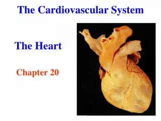
The Cardiovascular System
E N D
Presentation Transcript
The Cardiovascular System Ⅰ.Introduce Ⅰ) The organization — heart central pump — arteries carry blood away from the heart — capillaries — veins transport blood from the capillaries
The cardiovascular system Organization • Heart • A muscle pump to maintain the flow of blood, • Consist of four chambers (right and left atria, right and left ventricles) • Artery a.carry blood away from the heart • Veins v.carry blood back to the heart • Capillary microscopic vessels, the area of exchange between blood and tissue fluid
Ⅱ) Circuits 1.The systemic(greater) circulation left ventricle aorta arteries capillaries veins sup. and inf. vena cava right atrium
Here, the substance exchange between the blood and tissue cells through capillary wall takes place. The exchange includes oxygen, carbon dioxide, nutrients, water and inorganic ions, vitamins, hormones, metabolic products, antibodies, and defensive cells of various kinds. Then the arterial blood(rich in oxygen and nutrients) becomes venous blood(rich in carbon dioxide and metabolic products ) ,go through superior vena cava, inferior vena cava, coronary sinus, and the tributaries of them toward the right atrium. • Consequentially, this blood enters to the right ventricle.
Ⅱ) Circuits 2.The pulmonary (lesser) circulation right ventricle pulmonary trunk right and left pulmonary Arteries pulmonary capillaries(in lungs) pulmonary veins left atrium
blood gives off carbon dioxide and acquires oxygen. Thus, the venous blood becomes arterial blood, go through the pulmonary veins to the left atrium.
Vascular anastomosis When blood vessels connect to form a region of diffuse vascular supply it is called an anastomosis. Anastomoses provide critical alternative routes for blood to flow in case of blockages. • Anastomosis between a. • Anastomosis between v. • Arteriolovenular anastomosis • Collateral vessels Circulation
The Arteries Characteristics • Symmetry • In the trunk of the body consist parietal and visceral branches • Shortest possible course • Run on flexor surfaces • Usually do not pass directly through muscles, avoiding compression • Together with the veins and nerves in a sheath of fascia to form neurovascular bundle
Arteries of pulmonary circulation Pulmonary trunk Arises from right ventricle • Runs up, back ,and to the left • Bifurcates inferior to aortic arch into right and left pulmonary arteries, one for each lung
Arteries of thePulmonary Circulation Pulmonary trunk 1) left pulmonary a. arterial ligament A fetal vessel that connects the left pulmonary artery with the descending aorta and that normally closes at birth, becoming the arterial ligament 2) right pulmonary a.
Arteries of the Systematic Circulation 1. Aortic sinus 2. Ascending aorta 3. Aortic arch
Arteries of the Head and Neck A. Common carotid a. 1) external carotid a. a. superior thyroid a. b. lingual a. c. facial a.angular a. i. inferior labial a. ii. superior labial a.
Arteries of the Head and Neck d. superficial temporal a.e. maxillary a. i. middle meningeal a. ii. inferior alveolar a. iii. infraorbital a. f. occipital a. g. posterior auricular a.
Summary of Arteries of the Head • common carotid a. internal carotid a. brain • common carotid a. external carotid a. • 1. superior thyroid a. thyroid gland • 2. lingual a. tongue • 3. facial a. angular a. face • superior labial a. upper lip • inferior labial a. lower lip • 4.maxillary a. infraorbital a.middle face • & upper teeth • middle meningeal a. dura mater • inferior alveolar a. lower teeth • mental a. lower face • 5.superficial temporal a. temporal region • 6. posterior auricular a. posterior auricular region • 7. occipital a. posterior scalp • 8. ascending pharyngeal a. pharynx
Arteries of the Head and Neck 2) internal carotid a. a. carotid sinus: pressure receptor blood pressure b. carotid glomus:chemical receptor partial pressure of CO2 partial pressure of O2 hydrogen ion concentration, pH
Arteries of the Head and Neck B. Subclavian a. 1. Vertebral a. 2. Internal thoracic a. 1)superior epigastric a. 2)musculophrenic a. 3)anterior intercostal aa.4)pericardiacophrenic a.
Arteries of the Head and Neck 3. Thyrocervical trunk 1) inferior thyroid a. 2) transverse cervical a. 3) suprascapular a. 4. Costocervical trunk 5. Dorsal scapular a.
The Distribution of the Subclavian Artery subclavian a. 1. thyrocervical trunk inferior thyroid a. thyroid gland transverse cervical a. muscles of the back suprascapular a. muscles behind of the scapula 2.vertebral a. brain 3.internal thoracic a. superior epigastric a. upper abdominal wall musculophrenic a. pericardium & diaphragm anterior intercostal aa. intercostal space cardiacophrenic a. pericardium 4.costocervical trunk upper two costal space and adjacent muscles 5.dorsal scapular a. mm. of the back
Arteries of the Upper Limb A.Axillary a. 1. Superior thoracic a. pectoral muscles 2. Lateral thoracic a.serratus anterior and pectoral muscles 3. Thoracoacromial a. Pectoral branch acromial branch
Arteries of the Upper Limb 4. Subscapular a. 1) thoracodorsal a. latissimus dorsi 2) circumflex scapular a. infraspinatus 5. Posterior humeral circumflex a. deltoid muscle 6. Anterior humeral circumflex a. deltoid muscle
Arteries of the Upper Limb B. Brachial a. 1. Deep brachial a. triceps brachii& humerus 2. Superior ulnar collateral a. elbow joint 3. Inferior ulnar collateral a. elbow joint [first aid by pressing] [BP determination]
Arteries of the Upper Limb C. Radial a. 1. Radial recurrent a. 2. Superficial palmar branch superficial palmar arch 3. Principal a. of thumb
Arteries of the Upper Limb D. Ulnar a. 1. Ulnar recurrent a. 2. Common interosseous a. 1) anterior interosseous a. 2) posterior inter- osseous a. 3. Deep palmar branch
Arteries of the Upper Limb E. Superficial Palmar Arch Common palmar digital aa. proper palmar digital aa.
Arteries of the Upper Limb F. Deep Palmar Arch palmar metacarpal aa.
Summary of Arteries of the Upper Limb subclavian a. axillary a. 1. superior thoracic a. pectoral muscles 2. lateral thoracic a. serratus anterior and pectoral muscles 3.thoracoacromial a. muscles of thorax and scapular region 4.subscapular a. thoracodorsal a. latissimus dorsi circumflex scapular artery infraspinatus &subscapularis 5. post. humeral circumflex a. deltoid muscle 6.ant. humeral circumflex a. deltoid muscle
Summary of Arteries of the Upper Limb axillary a.brachial a. 1. deep brachial a. triceps brachii 2. superior ulnar collateral a. elbow joint 3. inferior ulnar collateral a. elbow joint 4. muscular branches anterior muscles of arm brachial a. radial a. 1. radial recurrent a. elbow joint 2.muscular branches anterior lateral muscles of forearm 3. principal artery of thumb thumb 4. superficial palmar branch superficial palmar arch fingers 5. deep palmar arch palmar metacarpal arteries common palmar digital arteries fingers
Summary of Arteries of the Upper Limb brachial a. ulnar a. 1. common interosseous a. posterior interosseous a. posterior muscles of forearm anterior interosseous a. anterior deep muscles of forearm 2. muscular branches anterior medial muscles of forearm 3. ulnar recurrent a. elbow joint 4.deep palmar branch deep palmar arch fingers 5. superficial palmar arch common palmar digital arteries proper palmar digital arteries fingers
Arteries of the Thorax Thoracic Aorta 1. Parietal branches 1) posterior intercostal aa. 2) subcostal a. 3) superior phrenic a.
Arteries of the Thorax 2. Visceral branches 1) bronchial a. 2) esophageal aa. 3) pericardial a.
Summary of Arteries of the Thorax aortic arch thoracic aorta Parietal branches 1. posterior intercostal aa. lower nine intercostal spaces 2. subcostal a. muscle of lower posterior thoracic wall 3. superior phrenic a. diaphragm Visceral branches 1. bronchial aa. bronchial tubes 2. esophageal aa. esophagus 3. pericardial a. pericardium
Arteries of the Abdomen A. Abdominal Aorta 1. Parietal branches 1) lumbar aa. 2) inferior phrenic a. • superior suprarenal aa. • 3) median sacral a.
Arteries of the Abdomen 2. Paired Visceral branches 1) middle suprarenal a. • 2) renal a. • a. inferior suprarenal a. b. segmental aa. of kidney 4) testicular a. (internal spermatic a.) ovarian a.
Arteries of the Abdomen 3) accessory renal a. 41.8%
Arteries of the Abdomen 3. Unpaired Visceral Branches 1) coeliac trunk a. left gastric a. b. common hepatic a. i. proper hepatic a. right gastric a. left branch right branch cystic a.
Arteries of the Abdomen ii. gastroduodenal a. right gastroepiploic a. superior pancreatico- duodenal a.
Arteries of the Abdomen c. splenic a. i. pancreatic branches ii. posterior gastric a. 60-80% iii. short gastric aa. iv. left gastroepiploic a. v. splenic branches
Arteries of the Abdomen 2)superior mesenteric a. i. inferior pancreatico-duodenal a. ii. jejunal aa. iii. ileal aa. iv. ileocolic a. appendicular a.
Arteries of the Abdomen v. right colic a. ascending colon vi. middle colic a. transverse colon
Arteries of the Abdomen 3)inferior mesenteric a. a. left colic a. descending colon b. sigmoid aa. sigmoid colon c. superior rectal a. upper rectum
Summary of Arteries of the Abdomen Parietal branches abdominal aorta 1. lumbar aa. posterior abdominal wall 2. inferior phrenic a. diaphragm superior suprarenal aa. suprarenal gland 3. median sacral a sacrum & coccyx 5th lumbar a. iliopsoas muscle Paired visceral branches abdominal aorta 1. middle suprarenal a. suprarenal gland 2. renal a kidney inferior suprarenal aa. suprarenal gland 3. internal spermatic a. testis or ovary
Summary of Arteries of the Abdomen Unpaired visceral branches abdominal aortacoeliac trunk 1. left gastric a.stomach 2.common hepatic a. proper hepatic a. left branch left liver right branch right liver cystic a gall bladder right gastric a stomach gastroduodenal a. right gastroepiploic a. right half of stomach & greater omentum superior pancreaticoduodenal a. pancreas & duodenum 3. splenic a. splenic branches spleen pancreatic branches pancreas short gastric aa. fundus of stomach left gastroepiploic a. left half of stomach & greater omentum
Summary of Arteries of the Abdomen abdominal aorta superior mesenteric a. 1. jejunal aa. jejunum 2. ileal aa. ileum 3. appendicular a. appendix 4. ileocolic a. ileum & caecum 5. right colic a. ascending colon 6. middle colic a. transverse colon abdominal aorta inferior mesenteric a. left colic a. descending colon sigmoid aa. sigmoid colon superior rectal a. upper rectum
Arteries of the Pelvis • Common Iliac a. • 1. Internal iliac a. Parietal branches 1) superior gluteal a. 2) inferior gluteal a. 3) obturator a. 4) iliolumbar a. 5) lateral sacral a.
Arteries of the Pelvis Visceral branches 1) umbilical a. superior vesical aa. 2) inferior vesical a. 3) inferior rectal a.
Arteries of the Pelvis 4) uterine a. 5) internal pudendal a.