Download
1 / 1
10 likes | 195 Views
Eberhard-Karls-Universität. UKT. Universitätsklinikum Tübingen Germany. Oral presentation March 7, 2002 9:15 a.m. Changes of the condylar morphology in patients wearing implant retained prosthodontics. IADR 80th General Session in San Diego, March 6-9, 2002

E N D
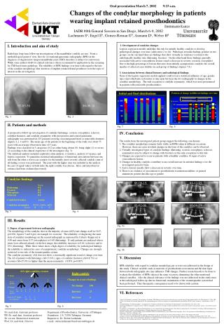
Eberhard-Karls-Universität UKT Universitätsklinikum Tübingen Germany Oral presentation March 7, 2002 9:15 a.m. Changes of the condylar morphology in patients wearing implant retained prosthodontics IADR 80th General Session in San Diego, March 6-9, 2002 Lachmann Sa, Engel Eb, Gomez-Roman Gb, Axmann Dc, Weber Hd I. Introduction and aim of study Radiologic long-term follow-up investigations of the mandibular condyle are rare. From a methodological point of view, the role of rotational panoramic radiography (RPR) in the diagnosis of degenerative temporomandibular joint (TMJ) disorders is subject to controversy. While some authors doubt its clinical relevance others recommend its application in the screening for TMJ hard tissue pathology. The reliability of RPR findings over time with regard to the fate of the condylar morphology after insertion of implant retained dental prostheses were the topic of interest in this investigation. 2. Development of condylar changes Logistic regression models underline this risk for initially healthy condyles to develop pathological changes over time (odds ratios 2 to 14).Pathologic articular findings polarize in two groups: on the one hand there are findings that show virtually no tendency to return to the radiologically healthy state (flattening, fracture). On the other hand those findings generally associated with active osteoarthrotic lesions trend to decrease in severity (erosion, osteophyte). Due to the high percentage of lesions that arise from initially asymptomatic condyles the overall number of affected cases doubles in each group of findings (figures 7,8.) 3. Associations between clinical features and radiological findings None of the logistic regression models applied could reveal a statistical influence of age, gender, type of prosthodontic restoration or time span between the two radiographs to changes in the condylar morphology. The only exception is condylar asymmetry, which was more pronounced in patients with removable prosthodontics. 97-05-30 98-06-16 Fig. 1 Fig. 2 II. Patients and methods A prospective follow-up investigation of condylar flattenings, erosion, osteophytes, sclerosis, condylar fractures, and condylar asymmetry with preoperative and control panoramic radiographs was performed in 223 patients receiving osseointegrated fixed or removable dentures (figures 1,2, 4 and 9). The mean age of the patients at the beginning of the study was about 55 years with an average observation time of 5 years. Findings were classified in 3 categories (0-2) as either being absent (0), being slight (1) or severe (2) according to the clinical experience of the investigator (fig. 3). The statistical analysis included explorative data analysis, к-statistics, analysis of variance and logistic regression. To guarantee statistical independence of functional associations between one side from the other a worst case scenario for the initially more severely affected condyle (sum of all finding scores) was performed. The joint with the higher sum was included in the analysis. In cases of equal values on both sides the right condyle was chosen. Intra- and interobserver variance had been evaluated previously. Fig. 7 Fig. 8 IV. Conclusion The results from the investigated patient group suggest the following conclusions: 1. The condylar morphology remains fairly stable on RPRs taken at different occasions. However, there are cases in which changes in the form of the condyles can be observed. 2. Virtually investigated types of condylar findings (flattening, erosion, osteophytes, sclerosis, asymmetry) may be subject to change with fractures as the only exceptions to this rule. 3. Changes are expected to occur in patients with a) healthy condyles, b) signs of active osteoarthrotic lesions. 4. Changes in healthy condyles contribute to an overall increase in articular findings over the investigated period of time. 5. Active lesions usually show a decrease in severity over time. 6. There is no evidence of associations to prosthodontic treatment modalities or general anamnestic patient data like age or gender. 88-08-19 93-12-15 95-02-06 00-06-19 Fig. 3 Fig. 4 III. Results 1. Degree of agreement between radiographs The morphology of the condyles does in the majority of cases (66%) not change at all (κ=0.47, p<0.001, figures 5 and 6 give an example for erosions). The reliability of diagnosing the same state of condylar morphology on the second radiograph as expressed by Cohen’s Kappa (κ) is 0.48 and varies from 0.37 (osteophytes) to 0.60 (flattening). If only patients are analyzed whose joints were affected already on the first image, the reliability increases to 0.61 (sclerosis) and to 0.91 (flattening). While these values show a high degree of reliability for pathological findings, the reliability is reduced in cases of asymptomatic condylar morphology. Changes are strongly restricted to one side of the stomatognathic system. The condylar asymmetry (AI) does not show a statistically significant trend of change over time. The AI of patients with flattenings (AI=13.8%), signs of condylar fractures (AI=18.7%) or erosions (AI=15.0%) is higher than the mean asymmetry (10.8%, p=0.007). Fig. 9a 88-08-19 93-12-15 95-02-06 00-06-19 Fig. 9b V. Discussion RPR reliability with regard to condylar morphology per se were not addressed in the design of this study. Clinical variables such as insertion of prosthodontic restorations and the time lapse between both radiographs also may influence TMJ changes. Further research needs to be done to evaluate the reliability of RPRs taken at the same occasion, eliminating the other mentioned clinical variables. Also the clinical relevance of the findings was not addressed in this study since at the radiological follow-up date no functional examination of the stomatognathic system had been performed. Thus therapeutic consequences need to be drawn with caution. VI. References 1. Engel E, Lachmann S, Axmann D. The prevalence of radiologic TMJ findings and self-reported orofacial pain in a patient group wearing implant dentures. Int J Prosthodont 14: 120-6, 2001. 2. Habets LL, Bezuur JN, Naeije M, Hansson TL. The orthopantomogram, an aid in the diagnosis of temporomandibular joint problems. II The vertical symmetry. J Oral Rehabil 15: 465-71, 1988. 3. John M, Pullinger AG. Pantomographie - ein diagnostisches Verfahren für knöcherne Veränderungen des Kiefergelenkes? Dtsch Zahnärztl Z 52: 553-7, 1997. 4. Könönen M, Kilpinen E. Comparison of three different radiographic methods in screening of temporomandibular joint involvements in patients with psoriatric arthritis. Acta Odont Scand 48: 271-7, 1990. 5. Lachmann S. Zur Häufigkeit von Kiefergelenkerkrankungen in einer zahnärztlichen Spezialsprechstunde. Eine klinisch-röntgenologische Systematik. In: Abteilung für Röntgendiagnostik in der Klinik für Zahn-, Mund- und Kieferkrankheiten. Hamburg-Eppendorf: Universität Hamburg, 2000. 6. Peltola J. Radiographic structural findings in the mandibular condyles of orthodontically treated children and young adults. Thesis. In: Departments of Dental Radiology, Paedodontics and Orthodontics, University of Helsinki. 1995. 7. Ruf S, Pancherz H. Is orthopantomography reliable for TMJ diagnosis? An experiment on a dry skull. J Orofacial Pain 9: 365-74, 1995. Reprint: Not in File 8. Türp JC, Vach W, Strub JR, Harbich K, Alt KW. Erkennung von mandibulären Asymmetrien auf der Panoramaschichtaufnahme. Schweiz Monatsschr Zahnmed 105: 755-9, 1995. 9. Engel E, Seeger P. Intra- und interindividuelle Reliabilität der bildgebenden Kiefergelenksdiagnostik. Eine Studie mit Panoramaschicht- und Magnetresonanztomographischen Aufnahmen von Patienten einer Funktionssprechstunde. Unpublished data, in preparation. Fig. 5 Fig. 6 aDr. med dent, Assistant professor bPD Dr. med dent, Assistant professor cDr. rer nat, Biometrical statistician dProf. Dr. med dent, Director Department of Prosthodontics, University of Tübingen Osianderstr. 2-8, 72076 Tübingen, Germany Requests to: Dr. Stefan Lachmann e-mail: stefan.lachmann@med.uni-tuebingen.de