Download
1 / 20
210 likes | 774 Views
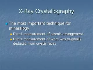
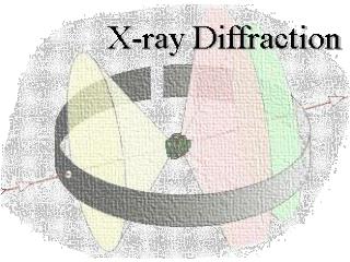
Structure of proteins by X-ray crystallography. George N. Phillips, Jr. ECE 532 Spring 2005. Points to be covered. What are proteins? How they are organized? How do we know their three-dimensional structures? Why do you need a crystal and how does one get a crystal?.

E N D
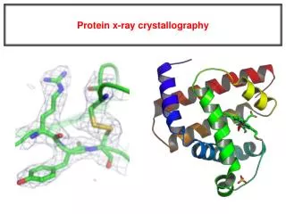
Structure of proteins by X-ray crystallography George N. Phillips, Jr. ECE 532 Spring 2005
Points to be covered • What are proteins? • How they are organized? • How do we know their three-dimensional structures? • Why do you need a crystal and how does one get a crystal?
Hierarchy in Protein Structure • Protein structure is usually described in terms of an organizational hierarchy : • Primary sequence • Secondary structure • Tertiary structure • Quaternary structure • But remember it is the sequence that controls all that follows!
Peptide Bond • The peptide bond influences all aspects of protein structure and function. • Key features: • 1. Planar • 2. Fairly rigid, due to partial double bond character. • 3. Almost always in trans configuration. • 4. Polar. Can form at least two hydrogen bonds. • 5. Places restrictions on the conformation of the polypeptide chain.
-helices This is the prototypical secondary structural element. It satisfies the hyrogen bonding requirements of the polypeptide chain (except for the ends). • Properties • It is compact and self contained. • Right handed • rise per residue, 1.5 Å • Residue per turn, 3.5Å • H-bond between C=O(n).....H-N(n+4) How do you define the start and end of an a-helix?
-sheets -sheets fulfill the hydrogen bonding potential of the main-chain atoms, except at the edges. Adjacent strands are usually close in sequence. Properties: Distance between Ca's is ~3.6 Å in an extended strand Distance between strands ~4.6 Å Strands are not flat. They have a characteristic right-handed twist How are sheets defined? Antiparallel b-sheet Parallel b-sheet
Turns Turns are a major structural component of proteins. Without them there could be no globular proteins. They are characterized by an irregular series of conformational angles that fold the chain back on itself. Turns are often very compact and well ordered, though they are hot-spots for evolution. Sometimes they are sites of flexibility, at other times they are quite rigid. Need to look carefully at the structure. Turns can be classified into a few well defined arrangements.
Major secondary structural elements of HIV-1 protease The secondary structure is mostly b-strand and random coil. There is one small helix and two b-turns. The b-sheets show their characteristic twist and associate into two layers, which is common in b-structures. The structure is compact, though this is not apparent in these ribbon representations.
Space Filling Representation of HIV-1 Protease Ribbon representations are convenient for showing the path of the polypeptide chain but are misleading with respect to the compactness. What is not evident in this representation? Hydrogens? Water? Dynamic properties?
Digression: Packing of a-helices and b-sheets in other proteins? • Secondary structural elements segregate into layers of helices, strands, and coil to optimize the packing of the side chains. Proteins do not have large holes on their interior, except for functional reasons.
Myoglobin and Hemoglobin Deliver Oxygen in Organisms with Vascular Systems
Light microscope Electron microscope X-ray Crystallography 2,000 X 1,000,000 x 100,000,000 X
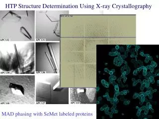
X-ray infrastructure Initial screening Optimization Identification George Phillips Craig Bingman Ed Bitto Simon Allard Jannelle Warrick Gary Wesenberg Determination
0 3 6 1 4 7 2 8 5