Download
1 / 10
100 likes | 242 Views
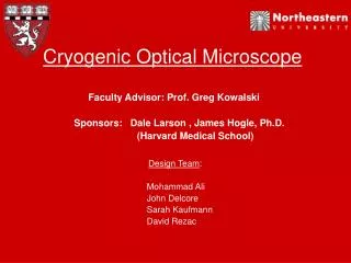
Observing Behavior at the Nanoscale using an Optical Microscope. Eann Patterson Composite Vehicle Research Center Michigan State University. Olympus IX70 Inverted Tissue Culture Microscope. 550nm with 45nm bandwidth. Diaphragm aperture. X60 objective (0.7 NA).

E N D
Observing Behavior at the Nanoscale using an Optical Microscope Eann Patterson Composite Vehicle Research Center Michigan State University
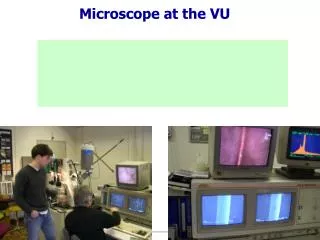
Olympus IX70 Inverted Tissue Culture Microscope 550nm with 45nm bandwidth Diaphragm aperture X60 objective (0.7 NA) high resolution (1324x1024) 16-bit monochrome cooled CCD camera
Resolving Nano-particles 100nm diasilica sphere ≡ 1/1000 human hair Diffraction Limit (220nm) 10mm dia (10,000nm) sphere ≡ 1/8 diameter of human hair wavelength of light (550nm) [Patterson & Whelan, Nanotechnology, 19(10)2008]
100nm silica particle in 3D condenser aperture diaphragm open condenser aperture diaphragm closed Composite images [Patterson & Whelan, Nanotechnology, 19(10)2008]
Brownian motion of 300nm particles Silica particles in ethanol: field of view 140x100mm
20000 nm Image of fibroblast cell in optical microscope using phase contrast with 100nm diameter nanoparticles
Image of fibroblast cell in optical microscope in nanoscope mode with 100nm diameter nanoparticles at t=0 20000 nm
Image of fibroblast cell in optical microscope nanoscope mode with 100nm diameter nanoparticles at t=100ms 20000 nm
Image of fibroblast cell in optical microscope nanoscope mode with 100nm diameter nanoparticles at t=100ms 5000nm/100ms ≡ 50 mm/s 2 m/hr 20000 nm
“Nothing tends so much to the advancement of knowledge as the application of a new instrument.” Elements of Chemical Philosophy (1812), in J. Davy (ed.), The Collected Works of Sir Humphry Davy(1839-40), Vol. 4, 37. Acknowledgements Professor Maurice P. Whelan