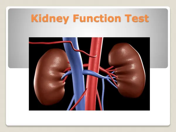
Kidney Function Tests
Kidney Function Tests. Main Functions of the Kidney. 1- Excretion of metabolic waste products & foreign chemicals 2- Regulation of water & électrolyte balance 3- Regulation of acid - base balance 4- Regulation of arterial blood pressure

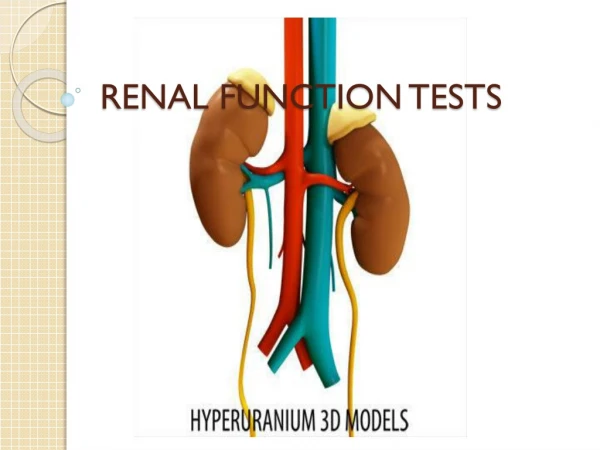
Kidney Function Tests
E N D
Presentation Transcript
Main Functions of the Kidney 1- Excretion of metabolic waste products & foreign chemicals 2- Regulation of water & électrolyte balance 3- Regulation of acid - base balance 4- Regulation of arterial blood pressure 5- Production of erythropoietin & activation of vitamin D 6- Other metabolic functions (as gluconeogenesis, etc..)
Renal Diseases • Overview • 1- Many renal diseases affect renal functions • 2- In some renal diseases, several functions are affected • 3- In other renal diseases, there is selective impairment of • glomerular function or one or more of the tubular functions • Most types of renal diseases causedestruction of • complete nephron
Major causes of renal diseases 1- Pre-renal diseases 2- Glomerular diseases 3- Tubular & interstitial diseases 4- Obstructive uropathies
Pre-renal diseases • The two major causes of reduced renal perfusion: Volume depletion (reduced volume of blood to the glomeruli) and/or relative hypotension • Prerenal disease is most commonly associated with anacute time course. • However, among patients with chronic kidney disease, the addition of a prerenal process may result in acute renal dysfunction
Glomerular diseases Causes: • Idiopathic • Secondary: neoplasia, autoimmune disease, drugs, infections, genetic • Two general patterns (with considerable overlap in some diseases) are seen: • Nephritic pattern Associated with inflammation on histological examination Urine: Active urine sediment with RBCs, WBCs, granular, red cell & other cellular casts Variable degree of proteinuria (mild to moderate in most cases). • Nephrotic pattern Not associated with inflammation on histological examination Urine: Proteinuria (moderate to severe . Most cases heavy proteinuria) An inactive urine sediment with few RBCs &WBCs cells or casts.
Tubular & interstitial diseases The tubular and interstitial diseases affecting the kidney can be divided into those that produce acute and chronic disease: • Acute tubulointerstitial disorders as acute tubular necrosis • Chronic tubulointerstitial disorders as polycystic kidney disease Obstructive Uropathy Obstruction to the flow of urine can occur anywhere from the renal pelvis to the urethra.
Nephrotic syndrome The nephrotic syndrome is caused by renal diseases that increase the permeability across the glomerular filtration barrier. It is classically characterized by four clinical features, but the first two are used diagnostically because the last two may not be seen in all patients. 1- Proteinuria: Urinary protein excretion greater than 50 mg/kg per day (heavy proteinuria) 2- Hypoalbuminaemia: Serum albumin concentration less than 3 g/dL (30 g/L) 3- Edema 4- Hyperlipidemia: increased cholesterol in blood Nephrotic syndrome is diagnosed by: Plasma Proteins Electrophoresis
Indications for assessing renal functions • Routine checkup • Older age • Chronic renal diseases • Decreased renal mass • Diabetes mellitus (DM) • Hypertension (HTN) • Autoimmune disease (as SLE, etc) • Systemic infections • Urinary tract infections (UTI) • Nephrolithiasis (renal stones) • Obstruction to the lower urinary tract (e.g. prostatic causes) • Drug toxicity
Assessment of Kidney Functions 1- Assessment of Glomerular Functions 2- Assessment of Tubular Functions
Biochemical Investigations of Glomerular Functions Assessment of glomerular filtration rate (GFR) is used an index of glomerular functions Measurement of GFR • Clearance Tests • Blood Creatinine • Blood Urea • Blood Uric Acid • Blood β2-microglobulin
Glomerular Filtration Is The first step in the production of urine Glomerular Filtration Rate (GFR) The amount of filtrate that flows out of all the renal corpuscles of both kidneys every minute In the normal adult, this rate is about 120 ml/minute i.e. about 180 liters / day GFR provides a useful index of the number of functioning glomeruli GFR can be estimated by measuring the urinary excretion of a substance that is completely filtered from the blood by the glomeruli and it is not secreted, not reabsorbed & not metabolized by the renal tubules.
Measurement of glomerular Filtration Rate • Clearance Tests: • Clearance is defined as the volume of plasma completely cleared from a • substance excreted in urine per minute • Normal Range: • About 110-120 ml/min in age of 20-40 years • Falls slowly & progressively to about 70 – 80 ml/min in ages over 80 years • More in males
U X V Clearance (ml/min) = __________________________________ P Measurement of glomerular Filtration Rate U is the concentration of substance in urine (in mmol/L) V is urine flow rate (in ml/min) P is the concentration of substance in bloodinmmol/L)
Measurement of glomerular Filtration Rate • Clearance Tests: • Accurate measurement of GFR by clearance tests requires determination of the • concentration in blood & urine of a substancethat is: • Freely filtered at glomeruli • Neither reabsorbed nor secreted by tubules. • Its concentration in plasma needs to remains constant throughout the period of • urine collection. • Better if the substance is present endogenously • Easily measured. • Creatinine meets most of these criteria
Measurement of glomerular Filtration Rate • Creatinine Clearance Test • Why Creatinine is used for testing clearance? • Creatinine is endogenously produced & is proportional to muscle mass • 1 to 2% of muscle creatine spontaneously converts to creatinine daily • Creatinine production is not affected by diet (no exogenous factors) • Creatinine is freely filtered at glomeruli at a constant rate. • Creatinine is not significantly reabsorbed by renal tubules • However, 10% of urinary creatinine is secreted by renal tubules (not • significant) • Blood levels of creatinine are maintained within narrow limits
Measurement of glomerular Filtration Rate Inulin clearance test Measurement of inulin clearance requires the infusion of inulin into the blood and is not suitable for routine clinical use Advantage of inulin clearance test over creatinine clearance test: Small quantity of creatinine is reabsorbed by the tubules and other quantities are actively secreted by the renal tubules So creatinine clearance is approximately 7% greater than inulin clearance. The difference is not significant when GFR is normal but when the GFR is low (less 10 ml/min), tubular secretion makes the major contribution to creatinine excretion and the creatinine clearance significantly overestimates the GFR (gives values greater than real ).
Plasma Creatinine Vs. Creatinine Clearance • Plasma creatinine correlates with GFR as does creatinine clearance in patients with • renal disease • Measurements of plasma creatinine are as effective in detecting early renal disease as • creatinine clearance • Plasma creatinine remains fairly constant throughout adult life while creatinine • clearance decreases with aging • Plasma creatinine measurements enable the progress of renal disease to be followed • with better accuracy than creatinine clearance • Creatinine Clearance is ONLY recommended (rather than serum creatinine) in: • Patients with early (minor) renal disease • Assessment of possible kidney donors • Detection of renal toxicity of some nephrotoxic drugs
Blood Urea • Urea is the major nitrogen-containing metabolic product of protein catabolism in humans. • Urea is filtered freely by the glomeruli • Plasma urea concentration is often used as an index of renal glomerular function • Non renal factors can affect the urea level (normal adults is level 5-39 mg/dl) as: • Mild dehydration • High protein diet (exogenous production factor) • Increased protein catabolism (as in Cushing`s disease, DM, starvation, thyrotoxicosis) • Reabsorption of blood proteins after a GIT hemorrhage • Accordingly, measurement of plasma creatinine provides a more accurate assessment than blood urea because there are many factors that affect urea level rather than renal causes
Blood Uric Acid • Renal handling of uric acid is complex and involves four sequential steps: • Filtration of virtually all the uric acid in capillary plasma entering the glomeruli • Reabsorptionin the proximal convoluted tubule of about 98 to 100% of filtered uric acid • Secretion of uric acid into the lumen of the distal portion of the proximal tubule • Further reabsorption in the distal tubule.
Blood Uric Acid cont. • In human, uric acid is the end product of the catabolism of the purine bases in in nucleic • acids (mainly DNA & RNA). • Approximately 75% of uric acid excreted is lost in the urine (remainder by GIT mainly) • Hyperuricemia • is defined by serum or plasma uric acid concentrations higher than 7.0 mg/dl in men or • greater than 6.0 mg/dl in women • Causes of hyperuricemia: • 1- Overproduction of uric acid: • Excessive intake of diets containing nucleic acids (esp. red meat). • Increased cellular breakdown (as in cancer, etc) • Genetic causes (as in Von Gierke`s Disease) • 2- Renal impairment (glomerular diseases) • SO YOU HAVE TO EXCLUDE OTHER RENAL CAUSES FOR HYPERURICEMIA
Plasma β2-microglobulin β2-microglobulin is: 1- A small protein 2- Present on the surface of most cells and in low concentrations in the plasma. 3- Completely filtered by the glomeruli & is reabsorbed & catabolized by proximal tubular cells. Results of measuring blood levels of β2-microglobulin: 1- Is a good index of GFR in normal people (as it is not affected by diet or muscle mass) 2- Since it is normally reabsorbed and catabolized in the tubules, β2-microglobulin blood level provides a sensitive method of assessing tubular functions. 3- BUT: It is increased in certain malignancies and inflammatory diseases.
Biochemical Investigations of Tubular Functionscont. Renal tubular functions is assessed by: • Urine Osmolality Measurements • Urine pH • Urine Volume • Urine Specific Gravity • Urine Appearance & Colour • Urine ProteinAmount • Urine Glucose Amount Measurement (Glucosuria) • Urine Amino Acids (Aminoaciduria)
Urine osmolality Osmolality: weight of solutes/ weight of solvent Urine osmolality: Concentration of all solutes (weight of all solutes / weight of urine) • Is a correct measure of the concentrating power of the kidney i.e. ability of the kidney to reabsorb water • Is done by determining the urine osmolality & then comparing this to the plasma. • Is highly affected by renal diseases. (the ability to concentrate the urine is affected ) So, urine osmolality serves as general marker of tubular function. Results of urine osmolality • If the urine osmolality is 600 mosm/kg or more, tubular function is usually regarded as intact • When the urine osmolality does not differ greatly from plasma (urine: plasma osmolality ratio=1), the renal tubules are not reabsorbing water (due to a tubular disease)
Urine osmolality cont. A patient with polyuria due to chronic renal failure is unable to produce either a dilute or concentrated urine Instead urine osmolality is generally within 50 mmol/kg of the plasma osmolality
Proteinuria • Normally: • Glomerular filtrate contains about 30mg/Litre protein; this corresponds to a total • filtered load of about 5g/24hours • Since only less than 200 mg protein is normally excreted in the urine each day, tubular • reabsorption must be very efficient
Proteinuria Normal protein amount n urine is < 200 mg/24hours urine collection Quantitative urine protein measurements should always be made on complete 24-hour urine collections. Types of proteinuria Glomerular proteinuria Tubular proteinuria Overflow proteinuria
Glomerular Proteinuria • Is caused by increased filtration of high molecular weight proteins (such as albumin) across the glomerular capillary wall. • Example: 1- Diabetic nephropathy 2- Nephrotic syndrome
Tubular Proteinuria • Overview Low molecular weight molecules such as smaller proteins (ß2-microglobulin, immunoglobulin light chains, retinol-binding protein ) & amino acids have molecular weights that are generally less than 25,000 in comparison to the 69,000 molecular weight of albumin. • Normally Smaller molecules (including smaller proteins & amino acids) can be filtered across the glomeruli & are then almost completely reabsorbed in the proximal tubule. • In tubular diseases: Interference with proximal tubular reabsorption can lead to increased excretion of these smaller proteins & amino acids (aminoaciduria) N.B. aminoaciduria due to inborn errors of amino acids metabolism must be excluded to diagnose tubular defects.
Overflow Proteinuria • Increased excretion of low molecular weight proteins can occur with marked overproduction of a particular protein, leading to increased glomerular filtration and excretion of this protein • This is due to (almost all causes): 1- Immunoglobulin light chains in multiple myeloma 2- lysozymesin acute myelomonocyticleukemia & in rhabdomyolysis 3- Hemoglobin in intravascular hemolysis