Kidney Function Testing
Kidney Function Testing. DR. Said Al ghora. An Introduction to the Urinary System. Transports urine towards bladder. Temporarily store urine. Produces urine. Conducts urine to exterior. The Function of Urinary System. A). Excretion & Elimination:

Kidney Function Testing
E N D
Presentation Transcript
Kidney Function Testing DR. Said Al ghora
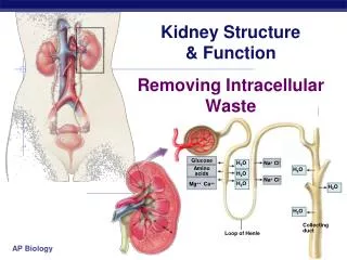
An Introduction to the Urinary System Transports urine towards bladder Temporarily store urine Produces urine Conducts urine to exterior
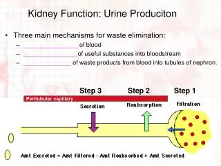
The Function of Urinary System A) • Excretion & Elimination: • removal of organic wastes products from body fluids (urea, creatinine, uric acid) • Homeostatic regulation: • Water -Salt Balance • Acid - base Balance • Enocrine function: • Hormones B) C)
Kidney – basic data • Urine excreted daily in adults:cca 1.5L • Kidney only ca 1% of total body weight, despite it • The renal blood flow= 20% of cardiac output • Plasma renal flow= PRF ca 600 mL/Min./1.73 M2 • Reflects two processes • Ultrafiltration (GFR): 180L/day • Reabsorption: >99% of the amount filtered
Renal threshold • Renal threshold of a substance is the concentration in blood beyond which it is excreted in urine • Renal threshold for glucose is 180mg/dL • Tubular maximum (Tm): maximum capacity of the kidneys to absorb a particular substance • Tm for glucose is 350 mg/min
Kidney Function • A plumbers view
How do you know it’s broken? • Decreased urine production • Clinical symptoms • Tests
Where can it break? • Pre-renal • Renal (intrarenal) • Post-renal (obstruction)
Causes of kidney functional disorders • Pre-renal e.g. decreased intravascular volum • Renal e.g. acute tubular necrosis • Postrenal e.g. ureteral obstruction
Signs and Symptoms of Renal Failure • Symptoms of Uraemia (nausea, vomiting, lethargy) • Disorders of Micturation (frequency, nocturia, dysuria) • Disorders of Urine volume (polyuria, oliguria, anuria) • Alterations in urine composition (haematuria, proteinuria, bacteriuria, leukocytouria, calculi) • Pain • Oedema (hypoalbuminaemia, salt and water retention)
Lab findings • Rising creatinine and urea • Rising potassium • Decreasing Hb • Acidosis • Hyponatraemia • Hypocalcaemia
Why Test Renal Function? • To identify renal dysfunction. • To diagnose renal disease. • To monitor disease progress. • To monitor response to treatment. • To assess changes in function that may impact on therapy (e.g. Digoxin, chemotherapy).
When should you assess renal function? • Older age • Family history of Chronic Kidney disease (CKD) • Decreased renal mass • Low birth weight • Diabetes Mellitus (DM) • Hypertension (HTN) • Autoimmune disease • Systemic infections • Urinary tract infections (UTI) • Nephrolithiasis • Obstruction to the lower urinary tract • Drug toxicity
Biochemical Tests of Renal Function • Measurement of GFR • Clearance tests • Plasma creatinine • Urea, uric acid and β2-microglobulin • Renal tubular function tests • Osmolality measurements • Specific proteinurea • Glycouria • Aminoaciduria • Urinalysis • Appearance • Specific gravity and osmolality • pH • osmolality • Glucose • Protein • Urinary sediments
Biochemical Tests of Renal Function • Measurement of GFR • Clearance tests • Plasma creatinine • Urea, uric acid and β2-microglobulin
Biochemical Tests of renal function In acute and chronic renal failure, there is effectively a loss of function of whole nephrons • Filtration is essential to the formation of urine tests of glomerular function are almost always required in the investigation and management of any patient with renal disease. • The most frequently used tests are those that assess either the GFR or the integrity of the glomerular filtration barrier.
Measurement of glomerular filtration rate GFR can be estimated by measuring the urinary excretion of a substance that is completely filtered from the blood by the glomeruli and it is not secreted, reabsorbed or metabolized by the renal tubules. • Clearance is defined as the (hypothetical) quantity of blood or plasma completely cleared of a substance per unit of time. • Clearance of substances that are filtered exclusively or predominantly by the glomeruli but neither reabsorbed nor secreted by other regions of the nephron can be used to measure GFR. • Inulin • The Volume of blood from which inulin is cleared or completely removed in one minute is known as the inulin clearance and is equal to the GFR. • Measurement of inulin clearance requires the infusion of inulin into the blood and is not suitable for routine clinical use V is not urine volume, it is urine flow rate
Biochemical Tests of Renal Function • Measurement of GFR • Clearance tests • Plasma creatinine • Urea, uric acid and β2-microglobulin
Creatinine • 1 to 2% of muscle creatine spontaneously converts to creatinine daily and released into body fluids at a constant rate. • Endogenous creatinine produced is proportional to muscle mass, it is a function of total muscle mass the production varies with age and sex • Dietary fluctuations of creatinine intake cause only minor variation in daily creatinine excretion of the same person. • Creatinine released into body fluids at a constant rate and its plasma levels maintained within narrow limits Creatinine clearance may be measured as an indicator of GFR.
Clinical Significance Elevated Creatinine is found in • Impaired renal function • Very high protein diet • Vary large muscle mass: body builders, giants, acromegaly patients • Rhabdomyolysis/crush injury • Drugs: • Trimethoprim • Amiloride
Clinical Significance • For renal transplant patients, an increase in serum creatinine of 2 mg/L has been used as a criterion of establishing rejection. • In other persons a change in creatinine of 2 mg/L would represent a 20% loss in renal function.
Specimen • One can analyze serum, plasma, or diluted urine. • The common anticoagulants (fluoride and heparin) do not cause interference, though heparin, which can be formulated as the ammonium salt, must be avoided in enzymatic methods that measure ammonia production. • Storage • 7 days at 4-25oC • At least 3 months at -20oC
Specimen • Urine should be diluted 1:100 • Bacterial contamination has been found to falsely lower creatinine values measured using the Jaffé reaction. • The mechanism of this interference appears to be bacterial production of a substance that retards the rate of the Jaffé reaction.
Enzymatic Method Creatinine aminohydrolase • Creatinine + H2O Creatine • Creatine +ATP Creatine-P + ADP • ADP + Phosphoenolpyruvate ATP + Pyruvate • Pyruvate + NADH Lactate + NAD+ • The difference in absorbance at fixed times during conversion is proportional to the concentration of creatinine in the sample Creatine Kinase Pyruvate Kinase Lactate dehydrogenase
Creatinine Clearance • Creatinine clearance is used to estimate the glomerular filtration rate (GFR). • One method of determining GFR from creatinine is to collect urine (usually for 24-hours) to determine the amount of creatinine that was removed from the blood over a given time interval. • Clearance is defined as the (hypothetical) quantity of blood or plasma completely cleared of a substance per unit of time. • The most frequently used clearance test is based on the measurement of creatinine. • Creatinine is chosen because it is freely filtered at the glomerulus and is not reabsorbed by the tubules.
However, a small amount of the creatinine (about 5%) in the final urine of healthy persons is derived from tubular secretion. • To do the test, one needs a precisely timed urine collection and a blood sample taken during the collection period. • Best results are obtained from a 24-h urine collection. • The test is initiated by having patients empty their bladder at the beginning of the timed period. • Urine is collected throughout the period, the bladder is again emptied at the end of the time period.
The 'clearance' of creatinine from plasma is directly related to the GFR if: • The urine volume is collected accurately • There are no ketones or heavy proteinuria present to interfere with the creatinine determination. • It should be noted that the GFR decline with age (to a greater extent in males than in females) and this must be taken into account when interpreting results.
Creatinine Clearance • Creatinine determinations are performed on both samples. The creatinine clearance is calculated from the following formula: • A person has a plasma creatinine concentration of 0.01 mg/ml and in 1 hour produces 60ml of urine with a creatinine concentration of 1.25 mg/mL.
Creatinine clearance (mL/min)= (UV)/P X 1.73/S • where U is urinary creatinine (mg/L), V is volume of urine (mL/min), P is plasma creatinine (mg/L), S is the calculated surface area of the patient, and 1.73 is the surface area (m2) of a standard 70 kg person. • The range of creatinine clearance in healthy persons corrected to a surface area of 1.73 m2 is 90 to 120 mL/min. • At low filtration rates, the creatinine clearance does not parallel true glomerular filtration rate because a relatively large portion of the urine creatinine is secreted rather than filtered.
Increased Muscle Mass Normal Muscle Mass Normal Muscle Mass Reduced Muscle Mass Creatinine Input Plasma Pool Content Kidney Output Normal Kidneys Diseased Kidneys Normal Kidneys Diseased Kidneys Effect of Muscle Mass on Serum Creatinine
Biochemical Tests of Renal Function • Measurement of GFR • Clearance tests • Plasma creatinine • Urea, uric acid and β2-microglobulin
Measurement of nonprotein nitrogen-containing compounds Catabolism of proteins and nucleic acids results in formation of so called nonprotein nitrogenous compounds. Protein Proteolysis, principally enzymatic Amino acids Transamination and oxidative deamination Ammonia Enzymatic synthesis in the “urea cycle” Urea
Plasma Urea Urea is the major nitrogen-containing metabolic product of protein catabolism in humans, • Its elimination in the urine represents the major route for nitrogen excretion. • More than 90% of urea is excreted through the kidneys, with losses through the GIT and skin • Urea is filtered freely by the glomeruli • Plasma urea concentration is often used as an index of renal glomerular function • Urea production is increased by a high protein intake and it is decreased in patients with a low protein intake or in patients with liver disease.
Plasma Urea • Many renal diseases with various glomerular, tubular, interstitial or vascular damage can cause an increase in plasma urea concentration. • The reference interval for serum urea of healthy adults is 5-39 mg/dl. Plasma concentrations also tend to be slightly higher in males than females. High protein diet causes significant increases in plasma urea concentrations and urinary excretion. • Measurement of plasma creatinine provides a more accurate assessment than urea because there are many factors that affect urea level. • Nonrenal factors can affect the urea level (normal adults is level 5-39 mg/dl) like: • Mild dehydration, • high protein diet, • increased protein catabolism, muscle wasting as in starvation, • reabsorption of blood proteins after a GIT haemorrhage, • treatment with cortisol or its synthetic analogous
Clinical Significance • States associated with elevated levels of urea in blood are referred to as uremia or azotemia. • Causes of urea plasma elevations: • Prerenal: renal hypoperfusion • Renal: acute tubular necrosis • Postrenal: obstruction of urinary flow
Increased protein catabolism: • Increased dietary protein • Severe tress: fever, etc • Rhabdomyolysis • Upper GI bleeding • Causes of urea plasma decrease • Decreased dietary protein • Increased protein synthesis ( Pregnant women , children ) • severe liver disease • Overhydration (IV fluids)
Specimen • Serum and heparinized plasma can be used for the urease/GLDH methods. • Fluoride will inhibit the urease reaction; therefore methods employing urease cannot use serum preserved with fluoride. • Ammonium heparin also cannot be used as an anticoagulant for urease methods. • Stability in serum or plasma: • 7 days at 4–8°C • 1 year at -20°C • Because of urea’s susceptibility to bacterial degradation, serum and urine samples should be kept at 4° to 8° C until analysis.
Urease/GLDH Method • The method is optimized for 2-point kinetic measurement. • Decrease in absorbance at 340 nm is proportional to concentration of urea
BUN / Creatinine Ratio • Normal BUN / Creatinine ratio is 10 – 20 to 1 • Creatinine is another NPN • Pre-renal increased BUN / Creat ratio • BUN is more susceptible to non-renal factors • Post-renal increased ratio BUN / Creat ratio • Both BUN and Creat are elevated • Renaldecreased BUN / Creat ratio • Low dietary protein or severe liver disease
Uric acid • In human, uric acid is the major product of the catabolism of the purine nucleosides, adenosine and guanosine. • Purines are derived from catabolism of dietary nucleic acid (nucleated cells, like meat) and from degradation of endogenous nucleic acids. • Overproduction of uric acid may result from increased synthesis of purine precursors. • In humans, approximately 75% of uric acid excreted is lost in the urine; most of the reminder is secreted into the GIT
Uric acid • Renal handling of uric acid is complex and involves four sequential steps: • Glomerular filtration of virtually all the uric acid in capillary plasma entering the glomerulus. • Reabsorption in the proximal convoluted tubule of about 98 to 100% of filtered uric acid. • Subsequent secretion of uric acid into the lumen of the distal portion of the proximal tubule. • Further reabsorption in the distal tubule. • Hyperuricemia is defined by serum or plasma uric acid concentrations higher than 7.0 mg/dl (0.42mmol/L) in men or greater than 6.0 mg/dl (0.36mmol/L) in women
Greater-than-normal levels of uric acid (hyperuricemia) may be due to: • Alcoholism • Diabetes • Gout • Hypoparathyroidism • Lead poisoning • Leukemia • Nephrolithiasis • Renal failure • Toxemia of pregnancy • Purine-rich diet • Excessive exercise • Chemotherapy-related side effects
Lower-than-normal levels of uric acid may be due to: • Fanconi syndrome • Wilson's disease • Syndrome of inappropriate antidiuretic hormone (SIADH) secretion • Multiple Sclerosis • Low purine diet
Gout • Gout is a kind of arthritis that occurs when uric acid builds up in the joints. • In Gout increased serum levels of uric acid lead to formation of monosodium urate crystals around the joints. • Acute gout is a painful condition that typically affects one joint. • Chronic gout is repeated episodes of pain and inflammation, which may involve more than one joint.
Specimen • Serum or plasma may be used; slight but insignificant positive bias (0.2 mg/dL) has been noted in plasma specimens as compared with serum. • Stability in serum / plasma: • 6 months at -20°C • 7 days at 4-8°C • 3 days at 20-25°C
Enzymatic Colorimetric Uricase • Uric acid + H2O + O2 Allantion + CO2 + H2O2 • TBHBA + 4- Aminoantipyrine + 2H2O2 Quinoneimine + 3 H2O • Uric acid is oxidized to allantoin by uricase. • The generated hydrogen peroxide reacts with 4-aminophenazone/ESPT to quinoneimine. POD
Metabolic features: Retention of: Urea & creatinine Na & water potassium with hyper-kalaemia Acid with metabolic acidosis Classification of Causes: Pre-renal reduced perfusion Renal inflammation infiltration toxicity Post-renal obstruction Acute Renal Failure
Reference Ranges • BUN 10 - 20 mg / dl • Creatinine 0.5 - 1.5 mg /dl • Uric Acid 3.0 - 7.0 mg / dl • Creatinine Clearance 90 - 130 ml / min • Ammonia 20 - 60 ug / dl • BUN / Creat Ratio 10 - 20 to 1