Download
1 / 30
331 likes | 918 Views
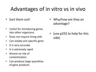
Advantages of in vitro vs in vivo. Sort them out! Useful for introducing genes into other organisms. Does not require living cells Can isolate one specific gene It is very accurate It is extremely rapid Almost no risk of contamination Can produce large quantities of gene products.

E N D
Advantages of in vitro vs in vivo • Sort them out! • Useful for introducing genes into other organisms. • Does not require living cells • Can isolate one specific gene • It is very accurate • It is extremely rapid • Almost no risk of contamination • Can produce large quantities of gene products • Why/how are they an advantage? • (use p255 to help for this side)
Summary sentence • In vivo is good for: • Accurate copies of a specific section of DNA to make recombinant DNA and/or making useful gene products • In vitro is good for: • Replicating DNA from small samples for analysis of crime scenes, blood samples for disease diagnosis, paternity suits, discover details about extinct organisms
Specification Medical diagnosis The use of labelled DNA probes and DNA hybridisation to locate specific genes. Once located, the base sequence of a gene can be determined by: • restriction mapping • DNA sequencing. Many human diseases result from mutated genes or from genes that are useful in one context but not in another, e.g. sickle cell anaemia. DNA sequencing and the PCR are used to produce DNA probes that can be used to screen patients for clinically important genes. The use of this information in genetic counselling, e.g. for parents who are both carriers of defective genes and, in the case of oncogenes, in deciding the best course of treatment for cancers. Candidates should understand the principles of these methods. They should be aware that methods are continuously updated and automated.
Locating and sequencing genes • DNA probes • DNA sequencing • Gel Electrophoresis • Restriction mapping
DNA Probes • DNA probes are simple, shortand single-stranded sections of DNA. • They will bindto complementary sectionsof other DNA strands. • Due to being labelled in some way, they make this ‘other DNA’ easily identifiable. Labelling with radioactivity Labelling with fluorescence
Question 6 (a) Strand of DNA; Short strand / up to 20 bases long; With base sequence that is complementary to part of target gene; Radioactive labelling / fluorescent labelling; 3 max (b) Identify carrier (of cancer gene); Identify which (cancer) gene present; Identify most effective treatment; 2 max Total 5
Learning Objectives • Recap how DNA probes and DNA hybridisation is used to locate specific genes. • Learn how the exact order of nucleotides on a strand of DNA can be determined. • Learn how restriction mapping can be used to determine nucleotide sequences.
Remember that probes can be used as an easy method of screening (detecting) for mutated genes.But also remember that the probe needs to be complementaryto the mutated gene.So this means, that to produce a probe, you first need to sequence your gene.How do we sequence genes?
Meet Frederick Sanger... • Biochemist • Cambridge University • English • Two Nobel Prizes • Still Alive Sanger’s work in the 1970’s, which earned him his second Nobel Prize, involved the sequencing of DNA. His method used modified nucleotidesthat do not allow another nucleotide to joinafter them in a sequence. Sanger Sequencing Method
Introducing Sanger Sequencing • The method is based on the premature endingof DNA synthesis. • If modified nucleotides are used during DNA synthesis, the process can be halted. What normally happens during DNA synthesis... T A T G G A T C T G A C C T T A G A T A C C T A G A C T G G A A T C What happens if you modify a nucleotide... You call these modified nucleotides, TERMINATORS T A T G G A T C A T A C C T A G A C T G G A A T C
What you need... • In Sanger Sequencing, four different terminators are used (A, C, T and G). • Due to this, four different reactions are run. • In each reaction, you have the following: • The DNA being sequenced. • A mixture of ‘normal’ nucleotides (A, T, C, D) • One typeof terminator nucleotide. • A primer. • DNA Polymerase. A T G C A C C
A A A A T T T T G G G G C T G A Remember that each tube probably contains millions of copies of the DNA template, countless nucleotides, and a good supply of the specific terminator nucleotide. Due to this, you get a variety of ‘partially completed’ DNA strands, because they have been ‘terminated’ at different points. A A A A C C C C C C C C
So what happens in each tube? • Now let’s imagine this is the sequence of the unknown DNA strand: • C C G T C T A G C A C T C A A G C T C T • Lets take the example of the tube with an adenine terminator What are the possible terminated sequences going to be when the reaction is over? A T G G G C A Because there are both ‘normal’ and ‘terminator’ nucleotides in the mixture, there is a chance that either is placed as the next base G G C A G A A G G C A G A T C G T G A A C G G C A G A T C G T G A G T T C G A C G G C A G A T C G T G A G T T C G A G A
Remember that this is happening in four test-tubes, each with a different type of terminator nucleotide.DNA fragments in each of the four tubes are going to be of varying lengths.Now the lengths of DNA need to be separated, so that we can see why we went through all of this trouble...
Gel Electrophoresis • When you’ve got a mess of DNA, especially DNA strands of varying lengths, you can separate them out using this technique. • The whole process relies on the fact that the phosphatesin the backbone of DNA, are negatively charged. • DNA fragments are placed in wellsat the top of an agar gel. • An electric current is applied over it. • Agar is actually a ‘mesh’, which resists the movement of the DNA fragments through it. • The DNA moves towards the positive electrode, but at different rates. • Small sections get there quicker.
Back to Sanger Sequencing • The fragments produced during the reactions can be separated using gel electrophoresis. • The smallest fragments will move furthest along the gel in a fixed period of time. • Due to being radioactively labelled, we can see where the DNA fragments end up, by placing photographic film over the gel, after the run. Terminator C Terminator A Terminator T Terminator G
Automated Sequencing • Nowadays, DNA sequencing is automated, using computers. • Nucleotides are fluorescently labelled with dyes. • Everything occurs in only a single tube. • And the separation can occur in one laneduring gel electrophoresis. Equipment used during the Human Genome Project.
Flash Video http://smcg.ccg.unam.mx/enp-unam/03-EstructuraDelGenoma/animaciones/secuencia.swf
Question 4 p282 • 4 a) polymerase chain reaction / PCR 1 • b) i) joins nucleotide together 1 • Not complementary bases. • ii) enables replication / sequencing to • start / keeps strands separate 1 • c) i) (modified nucleotide) does not form • bonds / react with other nucleotides; • does not ‘fit’ DNA polymerase / • enzyme / active site; • 1 max.
ii) AC 1 • Accept reading from right-hand side i.e. • TC. • d) i) different lengths / sizes / mass 1 • ii) radioactive primer 1 • iii) GAAGTCTCAG 1 • Accept reading from autoradiogram i.e. • CTTCAGAGTC.
Restriction Mapping • A process which allows the manipulation of a vector and can allow us to determine the sequence of nucleotides in a gene. • Can also be used to check that a piece of DNA is in the expected place within a vector.
Identification of RE sites • Use the sequence of plasmid pUC 12 to find the REs indicated at the bottom of the sheet. • Where in the sequence are they? Give the number of bases between each. • How can you map a plasmid? EcoRI 230 BamHI 248 PstI 270 HindIII 275 ScaI 1185
Back to pUC12 • If you cut with EcoRI, Pst1 and Sca1 what size fragments would you get? • Draw a restriction map and a diagram of the gel electrophoresis to show the results of this triple digest.
In summary so far...How can we... Reverse transcriptase Use of REs • produce DNA fragments? (2 ways) • Clone DNA? (2 ways) • Identify whether a gene has been taken up by the cell? (3 ways) • Identify smaller DNA sequences within a large sequence? • Sequence whole genomes or parts of DNA • Separate DNA fragments? • What has allowed all these processes to be sped up? (2 ways) In vivo (bacteria) PCR Enzyme markers Fluorescent markers Antibiotic markers Gene probe Sanger sequencing Gel electrophoresis Improvements in technology and automation
Q2 p280 AQA textbook • 2 a) i) transfer / carry genes from one organism to another / into bacteria /cells (1) • ii) cut open plasmid; • cut donor DNA, to remove gene /length of DNA; • cut donor DNA and plasmid with the same enzyme / enzyme that cuts at the same base sequence; • sticky ends / (overhanging) ends with, single strand / bases exposed; • association / attachment / pairing of complementary strand; 2 max. • iii) annealing / splicing / backbones joined / phosphodiester strand 1 • b) i) L and M 1 • ii) fragments 64 and 26 (kilobasesobtained) 1