Download

1 / 96
960 likes | 990 Views
Learn about pituitary gland structure & hormone secretion modes. Explore thyroid, parathyroid, pancreas, and adrenal gland histology. Identify gross structure & regions of equine pituitary.

E N D
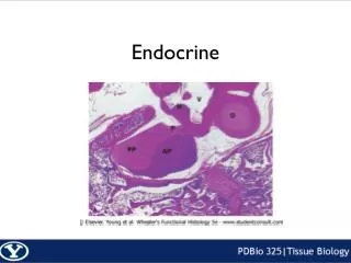
ENDOCRINE This resource is licensed under the Creative Commons Attribution Non-Commercial & No Derivative Works License
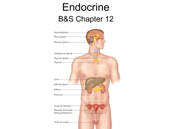
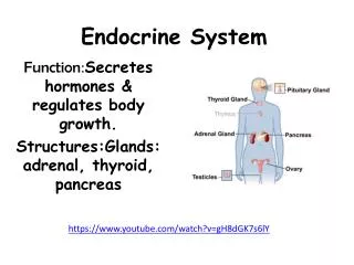
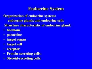
Objectives To describe the gross structure of the pituitary gland and be able to identify the pars nervosa, pars intermedia and pars distalis. Identify and describe the histological features of the pars nervosa and pars distalis and be able to relate these features to the different modes of hormone secretion. Identify and describe the histological features of the thyroid and parathyroid glands. Identify and describe the histological features of the pancreatic Islets of Langerhans. Identify and describe the histological features of the adrenal gland; in particular the different regions of the adrenal cortex.
Equine Head/Brain section locate Pituitary (Hypophysis)
Equine Head/Brain section locate Pituitary (Hypophysis) 1 Cerebrum 2 Optic nerve/chiasm 3 Pituitary (Hypophysis) 4 Mamillary body 5 Interthalamic adhesion 6 Aqueduct of Sylvius 1 1 corpus callosum fornix 5 cerebellum 6 2 4 3 pons medulla
SLIDE 159 Pituitary gland (hypophysis) At low magnification identify the main areas of the gland : A. B. A D B C 1.0 mm
SLIDE 159 Pituitary gland (hypophysis) At low magnification identify the main areas of the gland : A. Pars tuberalis. B. Pars distalis. C. Pars intermedia. D. Pars nervosa. A D B C 1.0 mm
SLIDE 159 Pituitary gland (hypophysis) At low magnification identify the main areas of the gland : A. Pars tuberalis. B. Pars distalis. C. Pars intermedia. D. Pars nervosa. A,B,C form the anterior hypophysis (adenohypophysis). D forms the posterior hypophysis (neurohypophysis). A D B C 1.0 mm
SLIDE 159 Pituitary gland (hypophysis) At low magnification identify the main areas of the gland : A. Pars tuberalis. B. Pars distalis. C. Pars intermedia. D. Pars nervosa. A,B,C form the anterior hypophysis (adenohypophysis). D forms the posterior hypophysis (neurohypophysis). From what germ cell layers does : B develop? A D B C 1.0 mm
SLIDE 159 Pituitary gland (hypophysis) At low magnification identify the main areas of the gland : A. Pars tuberalis. B. Pars distalis. C. Pars intermedia. D. Pars nervosa. A,B,C form the anterior hypophysis (adenohypophysis). D forms the posterior hypophysis (neurohypophysis). From what germ cell layers does : B develop? ectoderm. A D B C 1.0 mm
SLIDE 159 Pituitary gland (hypophysis) At low magnification identify the main areas of the gland : A. Pars tuberalis. B. Pars distalis. C. Pars intermedia. D. Pars nervosa. A,B,C form the anterior hypophysis (adenohypophysis). D forms the posterior hypophysis (neurohypophysis). From what germ cell layers does : B develop? ectoderm. D develop? A D B C 1.0 mm
SLIDE 159 Pituitary gland (hypophysis) At low magnification identify the main areas of the gland : A. Pars tuberalis. B. Pars distalis. C. Pars intermedia. D. Pars nervosa. A,B,C form the anterior hypophysis (adenohypophysis). D forms the posterior hypophysis (neurohypophysis). From what germ cell layers does : B develop? ectoderm. D develop? neuro-ectoderm. A D B C 1.0 mm
SLIDE 159 Pituitary gland (hypophysis) median eminence A : pars tuberalis B : pars distalis C : pars intermedia D : pars nervosa What are the principal secretory products of B,C and D? (The pars tuberalis contains secretory cells whose function is unknown). A D C B 1.0 mm
What are the principal secretory products of B,C and D? B : somatotropin, prolactin, FSH (follicle-stimulating hormone), LH (luteinizing hormone), TSH (thyroid-stimulating hormone), ACTH (adrenocorticotropic hormone). SLIDE 159 Pituitary gland (hypophysis) median eminence A : pars tuberalis B : pars distalis C : pars intermedia D : pars nervosa A D C B 1.0 mm
What are the principal secretory products of B,C and D? C : MSH (melanocyte-stimulating hormone). SLIDE 159 Pituitary gland (hypophysis) median eminence A : pars tuberalis B : pars distalis C : pars intermedia D : pars nervosa A D C B 1.0 mm
What are the principal secretory products of B,C and D? D : oxytocin, ADH (antidiuretic hormone or vasopressin). SLIDE 159 Pituitary gland (hypophysis) median eminence A : pars tuberalis B : pars distalis C : pars intermedia D : pars nervosa A D C B 1.0 mm
SLIDE 159 Pituitary gland (hypophysis) The pars distalis is separated from the pars intermedia. This space is the residual lumen of Rathke’s pouch (from which the anterior hypophysis develops) and is called the hypophysial cleft. The pars nervosa lies next to the pars intermedia. 250 µm
SLIDE 159 Pituitary gland (hypophysis) pars nervosa The pars distalis is separated from the pars intermedia. This space is the residual lumen of Rathke’s pouch (from which the anterior hypophysis develops) and is called the hypophysial cleft. The pars nervosa lies next to the pars intermedia. pars intermedia hypophysial cleft pars distalis 250 µm
SLIDE 159 Pituitary gland (hypophysis) Area of the pars distalis, identify : chromophilic cells a). acidophils (staining red). b). Basophils (pale-blue staining). chromophobes seen as clusters of nuclei. 25 µm
SLIDE 159 Pituitary gland (hypophysis) A : acidophils B : basophils C : chromophobes S : sinusoid A Area of the pars distalis, identify : chromophilic cells a). acidophils (staining red). b). Basophils (pale-blue staining). chromophobes seen as clusters of nuclei. S blood vessel B C S 25 µm
SLIDE 159 Pituitary gland (hypophysis) Area of the pars intermedia, identify : epithelioid cells 50 µm
SLIDE 159 Pituitary gland (hypophysis) epithelioid cells pars nervosa Area of the pars intermedia, identify : epithelioid cells pars intermedia pars distalis hypophyseal cleft (Rathke’s cleft) 50 µm
SLIDE 159 Pituitary gland (hypophysis) Area of the neurohypophysis, the pars nervosa. Identify : pituicytes (neuroglia-like cells). Herring bodies. 25 µm
SLIDE 159 Pituitary gland (hypophysis) H : Herring bodies P : pituicytes H Area of the neurohypophysis, the pars nervosa. Identify : pituicytes (neuroglia-like cells). Herring bodies. P H blood vessels H 25 µm
SLIDE 159 Pituitary gland (hypophysis) What are Herring bodies? 25 µm
What are Herring bodies? Accumulations of neurosecretions at the terminal regions of nerve fibres. Note the rich vasculature of this region. SLIDE 159 Pituitary gland (hypophysis) H : Herring bodies P P : pituicytes blood vessels H 25 µm
SLIDE 158 Pituitary gland (hypophysis) This section has been stained to show the chromophils and chromophobes of the pars distalis. Identify the main areas at this low magnification. 1.0 mm
SLIDE 158 Pituitary gland (hypophysis) infundibular stalk pars tuberalis pars intermedia This section has been stained to show the chromophils and chromophobes of the pars distalis. Identify the main areas at this low magnification. pars distalis pars nervosa This section shows a number of space artefacts, Probably produced during fixation of the tissue. The area indicated as hypophyseal cleft is much exaggerated and is shown better in section 159. hypophysial cleft 1.0 mm
SLIDE 158 Pituitary gland (hypophysis) Pars distalis : The chromophils have stained well, the acidophils staining pink and the basophils staining deep pink to purple. The chromophobes remain poorly stained . 25 µm
SLIDE 158 Pituitary gland (hypophysis) C A : acidophils B B : basophils Pars distalis : The chromophils have stained well, the acidophils staining pink and the basophils staining deep pink to purple. The chromophobes remain poorly stained . B C : chromophobes Bv : blood vessels Bv A A 25 µm
SLIDE 151 Thyroid gland Whole section viewed under low power. 1.0 mm
SLIDE 151 Thyroid gland trachea Whole section viewed under low power. thyroid gland lumen of trachea 1.0 mm
SLIDE 151 Thyroid gland Note the different sized follicles and the connective tissue capsule surrounding the gland. Connective tissue septa or trabeculae divide the gland into lobes. 250 µm
SLIDE 151 Thyroid gland connective tissue capsule thyroid follicles Note the different sized follicles and the connective tissue capsule surrounding the gland. Connective tissue septa or trabeculae divide the gland into lobes. connective tissue septum blood vessels in capsule 250 µm
SLIDE 151 Thyroid gland Thyroid viewed at higher power. What substance fills each follicle? 100 µm
SLIDE 151 Thyroid gland thyroid colloid the colloid fills the follicles space artefacts occur during histological fixation Thyroid viewed at higher power. What substance fills each follicle? Colloid; a suspension of iodinated thyroglobulin. A : artefact thyroid follicles A 100 µm
SLIDE 151 Thyroid gland thyroid colloid the colloid fills the follicles space artefacts occur during histological fixation Thyroid viewed at higher power. What substance fills each follicle? Colloid; a suspension of iodinated thyroglobulin. What is its function? A : artefact thyroid follicles A 100 µm
SLIDE 151 Thyroid gland thyroid colloid the colloid fills the follicles space artefacts occur during histological fixation Thyroid viewed at higher power. What substance fills each follicle? Colloid; a suspension of iodinated thyroglobulin. What is its function? Thyroglobulin is the storage form of thyroid hormones T3 and T4. A : artefact thyroid follicles A 100 µm
SLIDE 151 Thyroid gland Thyroid viewed at high power. What changes would be evident in a thyroid follicle under TSH stimulation? 50 µm
Thyroid viewed at high power. What changes would be evident in a thyroid follicle under TSH stimulation? The follicles would be smaller with less colloid. The cuboidal lining cells are taller, indicating active hormone synthesis and secretion. SLIDE 151 Thyroid gland colloid cuboidal epithelium lining follicles space artefact 50 µm
Thyroid viewed at high power. What changes would be evident in a thyroid follicle under TSH stimulation? The follicles would be smaller with less colloid. The cuboidal lining cells are taller, indicating active hormone synthesis and secretion. SLIDE 151 Thyroid gland colloid cuboidal epithelium lining follicles space artefact Follicles in an inactive thyroid are larger, filled with stored thyroid colloid and have a more flattened less active epithelium. 50 µm
SLIDE 151 Thyroid gland Where are ‘C’ cells located? 50 µm
Where are ‘C’ cells located? C cells also called parafollicular cells are derived from the neural crest; they occur singly amongst the follicle cells or more usually are found in small groups between the follicles. SLIDE 151 Thyroid gland C cells arrowed 50 µm
SLIDE 151 Thyroid gland What is the secretory product of C cells? 25 µm
What is the secretory product of C cells? Calcitonin : Secreted in response to high levels of blood calcium. It lowers these levels by reducing the resorption of bone. SLIDE 151 Thyroid gland C : C cells thyroid colloid C cuboidal follicular cells 25 µm
SLIDE 153 Parathyroid glands At low magnification note that these glands lie within the capsule of the thyroid. 1.0 mm
SLIDE 153 Parathyroid glands thyroid gland At low magnification note that these glands lie within the capsule of the thyroid. connective tissue capsule parathyroid 1.0 mm
SLIDE 153 Parathyroid glands The parenchymal cells form cords and are highly vascularised. What is the main cell type present in this gland? 250 µm
SLIDE 153 Parathyroid glands thyroid cords of principal cells The parenchymal cells form cords and are highly vascularised. What is the main cell type present in this gland? Principal cells (chief cells). blood vessels capsule 250 µm
SLIDE 153 Parathyroid glands What do the principal cells produce? 100 µm
What do the principal cells produce? Parathyroid hormone (PTH). SLIDE 153 Parathyroid glands capsule cords of principal cells blood vessel 100 µm