Download

1 / 17
170 likes | 199 Views
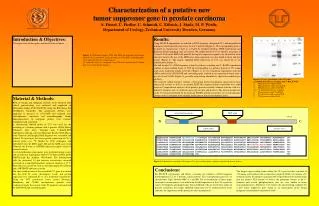
This article provides an overview of prostate carcinoma, one of the most common cancers worldwide. It discusses its incidence, risk factors, pathology, clinical features, diagnostic tests, and treatment options. The text language is English.

E N D
Incidence and epidemiology • One of the most common cancer in the world • risk factors • increasing age mostly occur in old man • race ( more common in black) • positive family history of cap • high dietary fat intake • smoking.
Pathology 95% of cap are adenocarcinomas rarely transitional cell ca. (more than 90% of the remaining 5%) *The diagnosis of cap differ from other tumor it depend on architecture. -70% of cap originate in the peripheral zone, -10-20% in the transitional zone, & -5-10% in the central zone.
*PIN (prostatic intraepithelial neoplasia) is a precursor for cap. *Usually the tumor is multifocal within the prostate with some variation in tumor grade. Grading Gleason grading system most commonly used, it relies upon the glandular architecture. There are 5 grades from well differentiated to undifferentiated glandular achitecture Gleason score by summation of the primary & secondary most common observed areas so its from 2 to 10 grades
Well differentiated tumours have Gleason sum of 2-4 • Moderately differentiated have Gleason sum of 5-6 • Poorly differentiated tumour have Gleason sum of 8-10 Gleason score of 7 called grey zone
Staging: (TNM staging system) Tx cannot assessed T0 no evidence of primary tumor Tis-carcinoma in situ (PIN) T1-discovered accidentally either by resected prostate or high PSA level. -T1a- less than 5% of resected tissue, -T1b- more than 5% of resected tissue, - -T1c- detected by elevated PSA T2-tumor palpable by DRE or visible by TRUS confined to prostate. T2a- confined to one lobe T2b- to both lobes T3-extracapsular extension including seminal vesicle. T4-tumor extend to bladder neck, rectum, or pelvic side wall.
Clinical features • Most cases with early stage Cap are asymptomatic • the presence of symptom suggest locally advance or metastatic disease. • Obstructive or irritative voiding symptoms • metastatic disease to the bone may cause bone pain • to spinal cord may cause spinal cord compression like parethesias and weakness of lower extremities and urinary or fecal incontinence • DRE may detect induration.
D.Dx of prostatic nodule include. • 1-Chronic granulomatous prostatitis, • 2-previous TURP or needle biopsy, • 3-prostatic calculus. • locally advance disease with lymphadenopathy may lead to lymphedema of lower limb.
Investigation • Uremia, if pt had obstructive uropathy. • Anemia, may be present in metastatic disease. • Tumor markers Alkaline phosphatase & serum acid phosphatase may be elevated, • PSA (prostate specific antigen) has great rule in diagnoses of cap • PSA: is serine protease produced by benign and malignant prostatic tissue so it organ specific but disease not specific • It is for screening and follow up treatment not for specific diagnosis of Cap • PSA normally up to 4ng/ml • PSA is vary with age,rase and prostatic volume
D.DX of high PAS 1-BPH 2-urethral instrumentation 3-infection 4-vigorous prostatic massage 5-prostatic biopsy or TURP But the elevation not significant. *PSA need about one month to return to its normal value after prostatic biopsy or TURP and only one week after prostatic message PSA velocity : PSA increased more than 0.75ng/ml/year is likely tumour PSA density ratio of PSA to gland volume if more than 0.1-0.15 biopsy indicated
Imaging. Like :- -TRUS (transrectal ultrasound), -Endorectal MRI, & -Bone scan (cap typically give osteoblastic lesion in bone). Prostatic biopsy :- Usually obtained under TRUS guidance. Indications 1- hard prostatic nodule by DRE 2-PSA more than 4 ng/ml and life expectancy more than 10 year
the most useful first-line test for the diagnosis of is prostate cancer is remain combination of DRE and PSA Treatment The treatment depend on -the grade & stage of the tumor, -the life expectancy of the pt, -associated morbidity, -the ability of therapy to ensure disease free survival, -the pt & physician preference.
A-Localized disease • Stage T1-T2 • 1- Watchful waiting and active surveillance • Watchfull waiting no thing is doing • in man with live expectancy of 10 years or less with • a- with very well-characterized early stage • b- low to intermediate grade cancer or less • Active surveillance based on • Serial and regular physical examinations • Serum PSA measurement • Repeat prostatic biopsy
2-Radical prostatectomy. Result depend on tumor stage &selection of better candidate pt usually with organ confined tumor. 3- Radiation thearpy 4-Radiation therapy& brachytherapy. improved imaging & the use of 3-dimention can increase the dose & decrease the toxicity to the surrounding normal organs. 5-Cryosurgery. Freezing of the prostate by using multiprobe cryosurgical device. Temperature may reach -25 to-50 C lead to tissue destruction.
The term brachytherapyrefers to a treatment technique that places radioactive sources in close proximity to or directl into the tumor. -can be classified as either interstitial or intracavity. *Interstitial brachytherapy involves the placement of radioactive needles, afterloaded needles or catheters, or radioactive seeds directly into the prostate, bladder, penis, or periurethral soft tissues. *Intracavitary brachytherapy includes placement of radioactive catheters into a lumen or orifice, such as in the urethra, to treat urethral and penile tumors. *Permanent implants involve the use of radioactive seeds that are left in the patient.
B-Metastatic disease. a single microscopic metastatic focus of prostate cancer in only one pelvic lymph nodes is a hallmark that its incurable by any currently available treatment modality Usually treated by endocrine therapy, because cap is hormonal dependant tumor about 70-80% of pt with metastatic cap responding to androgen deprivation. Complete deandrogenization is regarded as gold standard procedure need blockage of both testicular & adrenal androgen.
Testiculat androgen (95% of testosteron) can be blocked by either Surgical through bilateral orcheactomy or Medical by LHRH analogue which cause increase testosterone release in the first few weeks so should covered by flutamide to overcome flare up specially if there is spinal cord metastasis Andral androgen blocked by drug acting on the peripheral receptors like flutamide