Download
1 / 5
50 likes | 154 Views
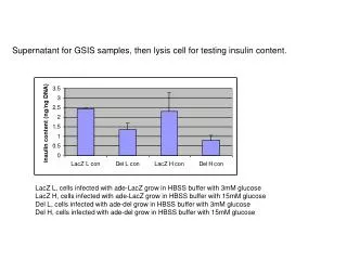
Supernatant for GSIS samples, then lysis cell for testing insulin content. LacZ L, cells infected with ade-LacZ grow in HBSS buffer with 3mM glucose LacZ H, cells infected with ade-LacZ grow in HBSS buffer with 15mM glucose Del L, cells infected with ade-del grow in HBSS buffer with 3mM glucose

E N D
Supernatant for GSIS samples, then lysis cell for testing insulin content. LacZ L, cells infected with ade-LacZ grow in HBSS buffer with 3mM glucose LacZ H, cells infected with ade-LacZ grow in HBSS buffer with 15mM glucose Del L, cells infected with ade-del grow in HBSS buffer with 3mM glucose Del H, cells infected with ade-del grow in HBSS buffer with 15mM glucose
. . . .. . . . . Granule fusion with membrane; Exocytosis of insulin 7 Microfilaments Secretory granules 5 Ca2+ Proinsulin to insulin; Packaging of insulin Microtubules 4 Golgi Transport of proinsulin to Golgi Glucose Transfer vesicles 3 Ca2+ contraction Of microfilaments Synthesis of preproinsulin; Excision of signal peptide; Disulfide bonds formed Endoplasmic 6 reticulum 2 mRNA production; Preproinsulin gene transcription Insulin storage 1 Nucleus Figure 2. Structural components (italicized) of the pancreatic -cell involved in glucose-induced biosynthesis and release of insulin. Biological structures
The defining property of KATP channels is the inhibition by intracellular ATP. KATP channels are octamers composed of four pore forming a -subunits (the inward rectifier channels Kir6.1 or Ki6.2) and four regulatory b -subunits (the sulphonylurea receptors SUR1 or SUR2. * My real-time PCR results proved sur.1 and sur6.2 RNA level in cells treated with ade-del C perk down 50%-25% compared to ade-LacZ. KATP channels are regulated by intracellular pH (24), protein kinase A (25,26) and protein kinase C. In addition, phosphatidylinositol 4,5 bisphosphate (PIP2), a membrane-bound phospholipid involved in G-protein-mediated signal transduction, can activate KATP channels .