Download
1 / 11
110 likes | 233 Views
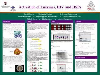
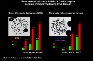
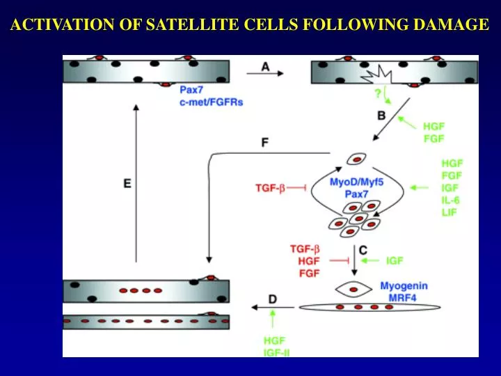
ACTIVATION OF SATELLITE CELLS FOLLOWING DAMAGE. CULTURE METHODS. 1. Muscle damage was induced by direct application of a 4-mm metal probe precooled in dry ice to the surface of the exposed muscle for 5 seconds. Muscles allowed to regenerate for 8-45 days.

E N D
CULTURE METHODS 1. Muscle damage was induced by direct application of a 4-mm metal probe precooled in dry ice to the surface of the exposed muscle for 5 seconds. Muscles allowed to regenerate for 8-45 days. 2. The tibialis anterior muscles was removed and weighed, and the tissue was dissociated using collagenase and dispase 3. The entire volume of the cell suspension generated from each muscle was plated in muscle medium(Ham’s F-10 media supplemented with 20% fetal bovine serum, 5 ng/ml bFGF, 100 U/ml penicillin G, and 100 g/ml streptomycin. 4. Differentiation was induced by plating cells on 6-well E-C-L (Upstate Biotechnology)–coated dishes and switching the media to a low serum DM in DME with either 2% horse serum or insulin-transferrin-selenium-A supplement.
Wild-Type CM Contains a Secreted Factor Involved in Myotube Growth Figure 1. (A) Bright field images of wild-type and NFATc2−/− myotubes after 24 and 48 hr in differentiation media (DM). Bar: 30 um. (B) NFATc2−/− myoblasts were induced to differentiate in DM or in wild-type conditioned media (CM) for 48 hr. Bar: 100 um. The number of nuclei in individual wild-type and NFATc2−/− myotubes was analyzed after incubation in DM or wild-type CM for 48 hr (bottom left). Wild-type and NFATc2−/− myotubes were treated with NFATc2−/− CM for 48 hr and analyzed as described above (bottom right). Data are mean ± standard error of three independent cell isolates. (*significantly different, p < 0.05).
IL-4 Is the Component of Wild-Type CM that Enhances Myotube Growth Figure 2. (A)NFATc2−/− myoblasts were treated with vehicle, 5 ng/ml IL-4, or 10 ng/ml IL-13 for 48 hr in differentiation media (DM). Bar: 60 um. Cells were treated with the indicated doses of IL-4 or IL-13 for 48 hr, and the myotubes were analyzed as in Figure 1B.(B) The DNA content of NFATc2−/− cells treated with vehicle or 5 ng/ml IL-4 was quantified after 48 hr in DM.(C) The percentage of nuclei within myotubes was calculated in NFATc2−/− cultures following treatment with vehicle or 5 ng/ml IL-4 at indicated times.(D) NFATc2−/− myoblasts were incubated in DM, wild-type conditioned media (CM), or wild-type CM treated with indicated doses of control IgG, IL-4, or IL-13 antibodies for 48 hr and were analyzed as in Figure 1B. Bar: 60 um. (E) NFATc2−/− myoblasts were treated with DM, wild-type CM, or IL-4−/− CM for 48 hr and analyzed as in Figure 1B. Data are mean ± standard error of three independent cell isolates (*significantly different from DM, p < 0.05).
NFATc2 Regulates IL-4 Expression in Skeletal Muscle Cells Figure 3. (A) IL-4 mRNA expression was examined by RT-PCR in wild-type and NFATc2−/− muscle cells after 24 hr in differentiated media (DM). Wild-type Th2 cells were included as a control. Representative ethidium bromide staining of agarose gel with three independent muscle cell isolates of each genotype is shown with 18S rRNA as an internal control.(B) The expression of IL-4 protein was analyzed in wild-type and NFATc2−/− CM by ELISA. Data are mean ± standard error of three independent cell isolates (*significantly different, p < 0.05).(C) NFATc2−/− myoblasts were infected either with control retrovirus (Ctrl) or with a retrovirus expressing recombinant NFATc2. IL-4 mRNA expression was analyzed by RT-PCR after 24 hr in DM. Representative ethidium bromide staining of agarose gel is shown with 18S rRNA as an internal control. Data are indicative of results from three independent experiments.
IL-4 Is Expressed by a Subset of Myotubes during Muscle Growth Figure 4. (A) Representative images of fusing cultures immunostained with an antibody against IL-4 after 24 or 36 hr in differentiation media (DM). Arrowheads indicate the same cell in both fluorescent and phase images. Bar: 60 um. The percentage of IL-4 positive cells was determined at the indicated times in three independent experiments.(B) Images of NFATc2−/− cells incubated with antibodies against IL-4 and wild-type cells incubated with secondary antibodies alone after 24 hr in differentiation media (DM).(C) After 24 hr in DM, wild-type cells were immunostained with an antibody against IL-4R and representative images are shown. Bar: 60 um.(D) At days 8 and 14 after injury, wild-type regenerating muscle sections were immunostained with an antibody against IL-4. Asterisks indicate the same myofibers in both fluorescent and haematoxilyn and eosin (H&E) stained images. Regenerating muscle sections at day 8 after injury from wild-type mice incubated with secondary antibodies alone and from IL-4−/− mice incubated with IL-4 antibodies are shown. Bar: 60 um.(E) mRNA expression for IL-4Ra, IL-13Ra1, IL-13Ra2, and c was examined by RT-PCR in cells after 0, 24, and 48 hr in DM. Macrophage mRNA was included as a control for IL-4R, IL-13R 1, and c, and mRNA from the glioblastoma cell line U251 was included as a control for IL-13R 2. Representative ethidium bromide staining of agarose gel is shown with 18S rRNA as an internal control. Data are indicative of results from three independent experiments.
Reduced Myofiber Size in IL-4−/− and IL-4R −/− TA and Soleus Muscles Is Muscle Cell Intrinsic Figure 5. (A) Representative sections of wild-type, IL-4−/−, and IL-4Ra−/− TA muscles are shown. Bar: 60 um. Data for myofiber cross-sectional areas (XSA) are mean ± standard error; N = 3 for wild-type, N = 7 for IL-4a−/−, and N = 4 for IL-4R−/−.(B) Frequency histograms showing the distribution of myofiber XSA in wild-type (n = 256), IL-4−/− (n = 731), and IL-4Ra−/− (n = 482) Tibialis anterior (TA) muscles (left) and wild-type (n = 447), IL-4−/− (n = 1060), and IL-4Ra−/− (n = 726) soleus muscles (right).(C) A representative wild-type myofiber immunostained with an antibody against dystrophin (red) and stained with DAPI (blue) illustrates the myonuclear number assay. The number of DAPI-stained nuclei within the dystrophin positive sarcolemma (arrow) were counted in soleus muscles from wild-type and IL-4−/−mice and expressed per 100 myofibers. Arrowhead indicates a nucleus outside the myofiber. Bar: 20 um. Data are mean ± standard error; N = 3 for each genotype.(D) The XSA of regenerating TA myofibers was determined at various time points after injury. Data are mean ± standard error; N = 4–7 for each genotype.(E) Wild-type, IL-4−/−, and IL-4Ra−/− myoblasts were induced to differentiate in differentiation media (DM) for 48 hr. Bar: 60 um. Myonuclear content was examined as in Figure 1B. Data are mean ± standard error of three independent cell isolates.(F) Wild-type or IL-4−/− myoblasts were infected with either a control retrovirus (Cntl) or a retrovirus expressing IL-4 (IL-4) and induced to differentiate for 48 hr. Bar: 30 um. Myotube cultures were analyzed as in Figure 1B. Data are mean ± standard error of three independent cell isolates (*indicates significantly different, p < 0.05).
IL-4 Acts on Myoblasts to Induce Myonuclear Accretion in Myotubes Figure 6. (A) Wild-type nascent myotubes (NMt) were cocultured for 24 hr in differentiation media (DM) with either wild-type or IL-4Ra−/− differentiated, mononucleated muscle cells (Mono). In addition, IL-4Ra−/− NMt were cocultured for 24 hr in DM with either wild-type or IL-4Ra−/− differentiated, mononucleated cells. Label colors represent the particular CellTracker dye used to stain each cell type. Bar: 60 um.(B) The percentage of myotubes containing dual label was determined and expressed as a percentage of the total myotubes analyzed (100). Data are the mean ± standard error of three independent cell isolates (*indicates significantly different, p < 0.05).
Model for the Role of IL-4 in the Recruitment of Myoblast Fusion during Muscle Growth Figure 7. Myoblast fusion occurs in two stages. In the first phase, a subset of differentiated myoblasts fuse together to form a nascent myotube with a limited number of nuclei. A second phase of myoblast fusion occurs with nascent myotubes. Under the control of NFATc2, IL-4 is secreted by nascent myotubes and induces this second phase of fusion through IL-4R on myoblasts. This action allows the accretion of nuclei within nascent myotubes and, along with protein accumulation, the formation of a large, mature myotube.
The NFATC2 pathway is critical in the second phase of myoblast fusion that occurs with nascent myotubes. A PGF2 activates NFATC2 in nascent myotubes through the FP receptor and promotes myoblast fusion and growth of nascent cells. B Under the control of NFATC2, IL-4 is secreted by nascent myotubes and induces the second phase of fusion through the IL-4R on myoblasts. This action allows the accretion of nuclei within nascent myotubes and along with protein accumulation, the formation of a large, mature myotube. Model for the NFATC2 pathway in the regulation of myoblast fusion.