Download

1 / 55
560 likes | 726 Views
Learn about the structure and function of joints and ligaments in the foot. Explore the muscles, retinacula, and movements associated with the ankle and metatarsophalangeal joints. Discover in-depth details on the arterial and nerve supply to the foot, emphasizing the importance of proper foot anatomy.

E N D
Contents of the anterior compartment • Muscles: • Tibialis anterior • Extensor digitorumlongus • Extensor hallucislongus • Peroneustertius • Anterior tibial artery • Deep peroneal nerve.
Retinacula • Deep fascia is thickened to form bands. • Retain tendons in place. • Superior Extensor Retinaculum • Inferior Extensor Retinaculum • Superior PeronealRetinaculum • Inferior PeronealRetinaculum
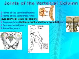
ANKLE or TALO-CRURAL JOINT Uni-axial synovial joint Modified hinge joint
Ligaments • Capsule • Synovial membrane • The ankle joint is bound medially by the strong • Deltoid ligament • Three lateral ligaments • •Anterior talofibular ligament • • Posterior talofibular ligament • • Calcaneofibular ligament
Deltoid ligament or Medial ligament Superficial part: • Anterior part or Tibio - navicular part • Intermediate part or Tibio - calcanean part • Posterior part or Posterior tibio - talar part Deep part/Anterior Tibiotalar
Attachments & parts of deltoid ligament Medial malleolus of Tibia Anterior Tibiotalar Tibiotalar Talus SustanticulumTali Tibionavicular Tibiocalcanean
Lateral ligaments Anterior Talofibular Lateral malleolus of Fibula Neck of Talus Posterior Talofibular Calcaneofibular ligament crossed by tendons of Peroneuslongus& Brevis
Movements of ankle joint Dorsiflexion: Close packed state. Position more stable. Plantar flexion: Loosely packed state of joint. Joint is less stable.
Movements Plantar Flexion: S1, S2 Gastrocnemius Soleus Assisted by Tibialis posterior, Flexor Digitorumlongus, Flexor Hallucislongus, Peroneuslongus and brevis in extreme plantar flexion
Dorsiflexion: L4, L5 Tibialis anterior Extensor Digitorumlongus, Extensor Hallucislongus, Peroneustertius
Arterial supply • Malleolar branches of Anterior Tibial Artery. • Malleolar branches of Peroneal Artery. NERVE SUPPLY • Branches from Deep Peroneal Nerve. • Branches from Tibial Nerve.
Interphalangeal joints Typical hinge joint Permit plantar flexion and dorsiflexion
Metatarso - phalangeal joints Ellipsoidal type of synovial joint. Ligaments: Capsule. Collateral ligaments. Deep metatarsal ligaments. Movements: Dorsi & plantar flexion. Adduction & abduction.
Tarso-metatarsal joints Plane synovial joints Gliding movements
Intertarsal joints SIX synovial cavities between tarsus and metatarsus : Sub-talar Talo-calcano-navicular Mid-tarsal / Transverse Calcaneo-cuboid tarsal • d) A joint cavity comprising cuneo-navicular, intercuneiform, cuneo-cuboid and intermediate and lateral cuneiform with bases of 2nd and 3rd metatarsal bone. • e) Between medial cuneiform and base of 1stmetarcarpal. • f) Between cuboid and 4th and 5th metacarpal
SUBTALAR JOINT /Posterior talo-calcanean joint Ligaments Capsule Medial and lateral talo-calcanean ligaments Interosseous talo-calcaneanligament Cervical ligament
Interosseous talo-calcanean ligament • Cervical ligament
Midtarsal or transverse tarsal joints TRANSVERSE TARSAL JOINT (MIDTARSAL JOINT) TALONAVICULAR JOINT CALCANEO-CUBOID JOINT
Midtarsal or transverse tarsal joint • Talonavicular joint • Calcaneocuboid joint Medial view of foot
Talo-calcaneo-navicular joint Restricted ball and socket type of joint Ligaments Talo-navicular ligament Plantar calcaneo-navicular Calcaneo-navicular part of bifurcated ligaments
The bifurcated ligament Y-shaped band. Its stem attached proximally to the anterior part of the upper calcanealsurface Distally it divides into 2 limbs - calcaneocuboid and calcaneonavicular parts. The calcaneocuboid ligament extends to the dorsomedial aspect of the cuboid The calcaneonavicular ligament is attached to the dorsolateral aspect of the navicular
Midtarsal or transverse tarsal joints • Talonavicular joint • Calcaneocuboid joint Lateral view of foot
CALCANEOCUBOID JOINT Saddle or sellar joint. Between anterior surface of calcaneus & posterior surface of cuboid CALCANEOCUBOID JOINT
Ligaments • Capsule • Calcaneocuboid part of bifurcate ligament • Long plantar ligament • Short plantar or Plantar calcaneocuboid ligament
BIFURCATE LIGAMENT DORSAL CALCANEOCUBOID LIGAMENT CALCANEOCUBOID JOINT
SHORT PLANTAR LIGAMENT (Plantar calcaneocuboid ligament) LONG PLANTAR LIGAMENT PLANTAR VIEW
Medial Lateral • The longestligament associated with the tarsus. • From Plantar surface of the calcaneus to plantar surface of thecuboid • The deep fibres are attached to the cuboid and more superficial fibres to the bases of the 2nd, 3rdand 4thand sometimes fifth, metatarsals. Long plantar ligament
Plantar calcaneocuboid ligament • Is deeper than the long plantar ligament • From the anterior calcaneal tubercle to • the cuboid
Movements INVERSION EVERSION
Inversion & Eversion These movements are essential to adjust the foot while walking on uneven surface.
Inversion Sole is directed downwards & medially. Associated with plantar flexion
Eversion Sole is directed downwards & laterally. Associated with dorsiflexion
Joints on which it occurs Subtalar joint Transverse tarsal joint. Talo-calcaneo-navicular joint Lateral view Medial view
Axis of movement Passes obliquely upwards, forward & medially from the heel end of calcaneusthrough sinus tarsi to dorso-medial surface of neck of Talus
Muscles causing Inversion Tibialis Anterior Tibialis Posterior
Muscles causing Eversion Peroneuslongus Peroneusbrevis Peroneustertius