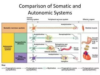
Somatic Motor Systems
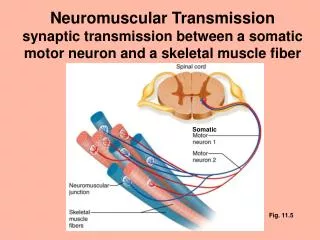
Somatic Motor Systems. Motor Units are the basic element of motor control in the somatic division. A motor unit consists of a single spinal or cranial motor neuron plus the skeletal muscle fibers that it innervates.

Somatic Motor Systems
E N D
Presentation Transcript
Motor Units are the basic element of motor control in the somatic division • A motor unit consists of a single spinal or cranial motor neuron plus the skeletal muscle fibers that it innervates. • Cell bodies of spinal somatic motor neurons are in lamina 10 of the spinal gray; cranial motor neurons are in nuclei of cranial nerves. • When a motor neuron is turned on, all of the muscle fibers it innervates are turned on.
Basic facts about mammalian skeletal muscle • Cells are large in diameter (up to 200 mu), and long (up to meters in larger animals) • Skeletal muscle is one of the two main forms of striated muscle: the contractile machinery is organized into the form of sarcomeres. • Each cell receives one and only one synapse from one and only one motorneuron. • Contractile activity is usually triggered by bursts of action potentials in motor neurons, which causes a smooth, sustained contraction called a tetanus. • There is no spontaneous contractile activity in the absence of input from motor neurons • Skeletal muscle is adapted by its structure for delivering maximal force at lengths close to its rest length.
Skeletal muscles fall into three functional classes • Type I slow twitch (oxidative) - this fiber type predominates in postural muscles. These fibers are characterized by smaller diameter, numerous mitochondria, little glycogen, numerous capillaries, and abundant myoglobin which gives them a red color. They can sustain contraction for long periods before fatiguing.
Fast twitch fibers are used at higher effort levels. • Type IIa fast twitch (oxidative) These fibers are used where moderate force must be sustained. They have a mixed metabolic strategy and intermediate fatigue properties. • Type IIb fast twitch (glycolytic) These fibers have the largest diameters and are characterized by few mitochondria, no myoglobin, few capillaries and large glycogen stores. They develop high forces but fatigue quickly.
Somatic units consist of single muscle fiber types • Smaller cell bodies are more easily brought to threshold; the smallest cell bodies belong to motorneurons that serve type I units, the next size category serves Type IIa units, and the largest sizes serve Type IIb units • As muscular effort is increased, additional units are recruited in the order • I IIa IIb • Probably in learned, high-speed activities, individual type II motor units in any individual muscle can be activated by higher centers through a motor program, without having to activate the type I fibers.
What kinds of synaptic inputs does a spinal motor neuron receive? • Sensory inputs from the ipsilateral body surface • Sensory inputs from muscle and joint receptors that activate stretch, withdrawal and tendon reflexes • Inputs from neurons in same or adjacent segments of the contralateral cord – for crossed reflex pathways • Descending pathways from higher motor centers that modulate spinal reflexes, regulate posture and apply motor programs for voluntary movements.
Spinal motorneurons integrate a large amount of information • The English neurophysiologist Sherrington described spinal motorneurons as “the final common path” of motor control to skeletal muscle. You could think of them as funnels with a wide mouth for collecting information (dendrites) but a narrow neck for delivering it to muscle fibers (axon).
Motor Control at the Spinal Level: The Stretch (Myotatic) Reflex What does it do? Activates motor units in a muscle in response to stretch of the muscle. Functions: • Maintains muscle length at a setpoint – particularly important for posture maintenance • Modulates force as needed to move muscle length to meet a new setpoint – important for load compensation in voluntary movements
Anatomical Components of the reflex arc • Sensors: muscle spindles (myotatic organs) containing tiny modified muscle fibers (intrafusal fibers) and two types of stretch receptor endings: Annulospiral endings (rapidly adapting) – associated with nuclear bag fibers Flower spray endings (slowly adapting) – associated with nuclear chain fibers • Afferents: Ia (largest size) axons connected to rapidly-adapting stretch receptors (annulospiral endings; II axons connected to slowly adapting receptors (flower spray endings) • Efferents: Alpha motor neurons to extrafusal fibers
Details of the muscle spindle Nuclear chain fiber Nuclear bag fiber Spindle capsule Flower spray ending Annulospiral ending Type II afferent Type Ia afferent Static gamma efferent Dynamic gamma efferent Gamma motorneurons are for resetting spindle length and are not within the stretch reflex arc Extrafusal fiber (for size comparison) Extrafusal fibers make up by far the greatest part of the muscle mass and provide all of the force. They are innervated by alpha motorneurons.
Spindle afferents are stimulated by muscle stretch • Stretching a muscle also stretches individual muscle spindles. The center of the spindle is the most mechanically compliant part, so that is where the most stretch occurs. This stretch activates the two kinds of afferents
The Stretch Reflex Arc The stretch reflex arc is monosynaptic; this is the only known monosynaptic reflex. All integration is carried out by the alpha motor neurons. the reciprocal inhibition of the opponent muscle is polysynaptic – there is at least one interneuron in the pathway These are alpha motor neurons
Stretch reflexes are the power steering of voluntary movements • Fact 1: the intrafusal fibers cannot shorten the spindle unless the muscle also shortens • Fact 2: spindle shortening and muscle shortening must match each other. • If muscle shortens and spindle doesn’t, the spindle becomes slack and unresponsive. • If spindle is activated and shortens but muscle is also activated and doesn’t shorten, the center part of the spindle gets stretched and the afferents are stimulated.
Steps in a reflex-assisted voluntary movement: • 1. Higher motor centers send descending signals that activate particular muscle groups – this is believed to involve coactivation of alphas to initiate muscle activity and gammas to reset length setpoint of spindles. • 2. Question: Is muscle at new setpoint length yet? • YES: activity in Ia and II afferents from muscle spindles is at baseline level – no additional motor unit recruitment • NO: activity in spindle afferents is above baseline – spindle inputs and descending central inputs summate on alphas, recruiting additional motor units.
The Tendon Reflex Arc The Golgi tendon organ reflex is believed to mediate overload protection by inhibiting the motor units of the stressed muscle and activating those of the opponent muscle. The axons of Golgi tendon organs are Type II afferents
The Withdrawal Reflex Arc The flexion or withdrawal reflex mediates protective withdrawal of an injured appendage. The crossed extension component of the reflex increases activity in extensors of the contralateral limb when the flexion reflex is activated.
What can a spinal animal do? (Cord is transected at a level just below the medulla) • 1. generate muscle tone if alpha motor neurons are modulated at correct level – but this generally doesn’t happen if cord is transected below the medulla. • 2. generate alternate stepping movements if the animal is supported – due to mutual inhibitory connections between extensors and flexors and between opposite limbs – in fact such animals can run on a treadmill, changing gaits as the treadmill is speeded up, in a way indistinguishable from intact animals. http://www.youtube.com/watch?v=wPiLLplofYw • 3. perform spinal reflexes (withdrawal, genitourinary reflexes) – but in human patients reflex responsiveness depends very much on the management of the patient after the injury. • No voluntary movements are possible for myotomes below the level of section.
Higher Somatic Motor Control From the Vestibular Nucleus of the Brainstem Upward
Higher Motor Control is distributed between 4 interacting brain areas • Brainstem – antigravity reflexes, including responses to vestibular inputs • Motor Cortex – distal muscles of appendages for highly controlled manipulative movements • Cerebellum – coordination of rapid movements • Basal Ganglia – execution of motor programs involving multiple large muscle groups • The interneurons in these areas are sometimes called upper motor neurons, in contrast to spinal motor neurons, which are called lower motorneurons.
Vestibular System • Components: Semicircular canals: responsive to rotational acceleration of head relative to the inertial frame of reference Saccule and utricle: detect head position relative to pull of gravity and linear acceleration of head
Semicircular canals detect head rotation relative to the inertial frame of reference endolymph lags behind as head rotates, displacing barrier and stimulating hair cells endolymph Gelatinous barrier Hair cells Branch of vestibular nerve
The two sets of 3 canals are oriented to detect head rotation in 3 planes
The saccule and utricle (one set on each side of the head) detect head position relative to gravity, linear acceleration, and low-frequency vibration Acceleration in any direction creates a direction and magnitude specific pattern of stimulation of the hair cells Gelatinous filling Calcium carbonate weight Filling displaced in opposite direction Head acceleration Branch of vestibular nerve
Vestibular System and brainstem postural reflexes • Brainstem modulates outflow to extensors to maintain postural setpoints, particularly in response to changes in head position. • Deviations relative to pull of gravity are detected by muscle/joint receptors and the vestibular system, activating righting responses. • Vestibulo-ocular and vestibulo-collic reflexes allow you to fix your gaze on a target irrespective of head and body movements
Midbrain transection creates a decerebrate animal • One classic sign of damage to pathways between medulla and more rostral centers is a tonic “decerebrate rigidity”. This arises from the fact that the vestibular nuclei of the brainstem mostly provide excitatory input to spinal motor neurons that serve extensors, whereas the higher centers provide some mixture of excitatory and inhibitory input. When the inhibitory component is removed, excitatory inputs predominate, so appendages are rigidly extended.
Like the primary somatosensory cortex, the primary motor cortex has a somatotopic organization The motor cortical homunculus
Cortical columns in motor cortex Within the motor cortex, individual columns consist of cells that are involved in controlling motion around a single joint. Within a column, there are cells that become active for specific angles of movement of that joint. As a result of this, the motor cortex is particularly important for finely controlled manipulations in primates. It is less important in animals that do not use their appendages for manipulation.
The cerebellum is involved in predicting and controlling the future position of rapidly moving body parts • Decomposition of complex movements is a symptom of cerebellar damage • Cerebellar neurons only interact with other brain motor areas and do not connect directly with spinal motor neurons.
The basal ‘ganglia’ include 3 interior nuclei • Globus pallidus • Caudate nucleus • Putamen
Athetosis – writhing movements Ballisms – flinging movements Chorea – ‘dance-like’ movements Parkinson’s disease – rigidity and difficulty in initiating movements In many cases these seem to involve inappropriate playing out of motor programs Parkinson’s disease involves death of dopaminergic neurons in the substantia nigra – dopamine is an important modulator of neuronal activity in the putamen. Basal ganglion diseases cause uncontrolled movement, suggesting that motor programs are organized and stored primarily by the basal ganglia
Two main kinds of pathways lead from brain motor areas down to the spinal cord • 1. Pyramidal tract = direct corticospinal tract Axons coming from pyramidal cells in the motor cortex pass through the thalamus, decussate in the medullary pyramids and descend in lateral corticospinal tract in cord – these axons mainly serve distal muscles that are involved in reaching and manipulation.
Pyramidal and ventromedial tracts The ventromedial tract carries a small number of fibers that run direct from the motor cortex to the spinal cord – these mostly innervate axial muscles.
Extrapyramidal Pathways • 2. The extrapyramidal tracts contain axons emanating from the basal ganglia and brainstem nuclei – some decussate in the brainstem – some don’t decussate at all (!). In contrast to the direct corticospinal tract, these are polysynaptic pathways. These pathways are mainly involved in control of axial and girdle muscles. They are mainly involved in movements that can occur without conscious control • Tectospinal – control head movements, reflexive responses to visual and auditory threats, including escape responses. • Vestibulospinal – modulate stretch reflexes, match posture to head position • Rubrospinal- an additional pathway from motor cortex to spinal motorneurons - innervates mainly flexors – so works together with vestibulospinal outputs to regulate posture • Reticulospinal – from reticular formation – adjust posture to reflect alertness
What can a decerebrate animal do? • Brain is transected above midbrain but below thalamus • Righting reflexes – driven by vestibular system – but not those driven by visual system • Static responses – modifications of posture in response to the needs of a particular situation – eg tonic neck responses
What can a decorticate (high midbrain transection) animal do? • Nonprimates (rats, cats): walk, eat, drink, copulate, raise pups (but not make a nest), learn to some extent (eg, conditioned reflexes), show “sham” emotion, respond to touch, pain and sound • decorticate cats attack moving objects – even though they can’t see. • Primates – decortication causes very serious global disability – one of the ways we know about this is from anencephalic children, who in almost all cases enter a chronic vegetative state.
Diseases of central motor pathways • “Upper motor neuron” lesions (i.e. in cerebral white matter that interrupt descending pathways): paralysis on contralateral side, increased muscle tone and stretch reflex, + Babinski sign and other infantile reflexes reappear. • Basal nuclei diseases: Parkinson’s D., Huntington’s chorea – impoverished movement repertoire (bradykinesia) http://www.youtube.com/watch?v=0E7x1mPa3iM&feature=related http://www.youtube.com/watch?v=OveGZdZ_sVs&feature=related • Corticospinal tract lesions (i.e. in spinal cord) paralysis on ipsilateral side, decreased muscle tone and reflex strength; muscle wasting. • “Lower motor neuron disease” (i.e. lesions to single spinal segments): paralysis of specific muscle groups, decreased muscle tone; muscle wasting.